Christoph Trenzinger, Caroline Kopittke, Barbora Kalousková, Nemanja Šikanić, Marina Bishara, Gerhard J. Schütz and Mario Brameshuber
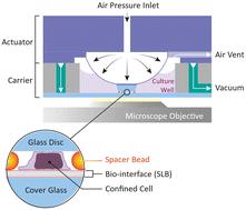
{"title":"在功能化生物界面上限制t细胞的微型装置。","authors":"Christoph Trenzinger, Caroline Kopittke, Barbora Kalousková, Nemanja Šikanić, Marina Bishara, Gerhard J. Schütz and Mario Brameshuber","doi":"10.1039/D5LC00248F","DOIUrl":null,"url":null,"abstract":"<p >Mechanical stimuli are an integral part of the natural cellular microenvironment, influencing cell growth, differentiation, and survival, particularly in mechanically challenging environments like tumors. These stimuli are also crucial in the T-cell microenvironment, where they play a role in antigen recognition and pathogen detection. To study T-cell mechanobiology effectively, <em>in vitro</em> methods must replicate these mechanical stimuli induced by compression, tension or shear flow, in the presence of antigen-presenting cells (APCs). While custom-made microdevices and microfluidic chips have successfully observed bulk cell behavior under mechanical strain, no existing device fully replicated the T-cell mechanoenvironment comprehensively. In this study, we developed a microdevice that integrates the mechanoenvironmental aspects of an APC mimicry with compression under live-cell imaging conditions. This device allows for precise confinement of cells between two glass surfaces, which can be individually coated with functional bio-interfaces. The microdevice is reusable and enables presetting of confinement heights, manual seeding of cells and the assembly of components directly at the microscope. To validate our microdevice we confined primary mouse T-cells on different APC-mimicking supported lipid bilayers while monitoring their morphology and migratory behaviour over time. To study the effect of confinement on TCR signalling, we tracked intracellular calcium levels and quantified Erk1/2 phosphorylation by immunostaining. We observed that T-cell morphology and motility are affected by confinement but also by bilayer composition. Moreover our findings suggest that confinement, despite not interfering with T-cell activation, might increase TCR background signalling in resting T-cells. Importantly, our microdevice is not limited to T-cell research; it can also serve as a platform for studying mechanical stimulation in other cell types, cell aggregates like spheroids and organoids, or even tissue samples in the presence of various bio-interfaces.</p>","PeriodicalId":85,"journal":{"name":"Lab on a Chip","volume":" 11","pages":" 2654-2668"},"PeriodicalIF":5.4000,"publicationDate":"2025-04-22","publicationTypes":"Journal Article","fieldsOfStudy":null,"isOpenAccess":false,"openAccessPdf":"https://pubs.rsc.org/en/content/articlepdf/2025/lc/d5lc00248f?page=search","citationCount":"0","resultStr":"{\"title\":\"Microdevice for confinement of T-cells on functionalized bio-interfaces†\",\"authors\":\"Christoph Trenzinger, Caroline Kopittke, Barbora Kalousková, Nemanja Šikanić, Marina Bishara, Gerhard J. Schütz and Mario Brameshuber\",\"doi\":\"10.1039/D5LC00248F\",\"DOIUrl\":null,\"url\":null,\"abstract\":\"<p >Mechanical stimuli are an integral part of the natural cellular microenvironment, influencing cell growth, differentiation, and survival, particularly in mechanically challenging environments like tumors. These stimuli are also crucial in the T-cell microenvironment, where they play a role in antigen recognition and pathogen detection. To study T-cell mechanobiology effectively, <em>in vitro</em> methods must replicate these mechanical stimuli induced by compression, tension or shear flow, in the presence of antigen-presenting cells (APCs). While custom-made microdevices and microfluidic chips have successfully observed bulk cell behavior under mechanical strain, no existing device fully replicated the T-cell mechanoenvironment comprehensively. In this study, we developed a microdevice that integrates the mechanoenvironmental aspects of an APC mimicry with compression under live-cell imaging conditions. This device allows for precise confinement of cells between two glass surfaces, which can be individually coated with functional bio-interfaces. The microdevice is reusable and enables presetting of confinement heights, manual seeding of cells and the assembly of components directly at the microscope. To validate our microdevice we confined primary mouse T-cells on different APC-mimicking supported lipid bilayers while monitoring their morphology and migratory behaviour over time. To study the effect of confinement on TCR signalling, we tracked intracellular calcium levels and quantified Erk1/2 phosphorylation by immunostaining. We observed that T-cell morphology and motility are affected by confinement but also by bilayer composition. Moreover our findings suggest that confinement, despite not interfering with T-cell activation, might increase TCR background signalling in resting T-cells. Importantly, our microdevice is not limited to T-cell research; it can also serve as a platform for studying mechanical stimulation in other cell types, cell aggregates like spheroids and organoids, or even tissue samples in the presence of various bio-interfaces.</p>\",\"PeriodicalId\":85,\"journal\":{\"name\":\"Lab on a Chip\",\"volume\":\" 11\",\"pages\":\" 2654-2668\"},\"PeriodicalIF\":5.4000,\"publicationDate\":\"2025-04-22\",\"publicationTypes\":\"Journal Article\",\"fieldsOfStudy\":null,\"isOpenAccess\":false,\"openAccessPdf\":\"https://pubs.rsc.org/en/content/articlepdf/2025/lc/d5lc00248f?page=search\",\"citationCount\":\"0\",\"resultStr\":null,\"platform\":\"Semanticscholar\",\"paperid\":null,\"PeriodicalName\":\"Lab on a Chip\",\"FirstCategoryId\":\"5\",\"ListUrlMain\":\"https://pubs.rsc.org/en/content/articlelanding/2025/lc/d5lc00248f\",\"RegionNum\":2,\"RegionCategory\":\"工程技术\",\"ArticlePicture\":[],\"TitleCN\":null,\"AbstractTextCN\":null,\"PMCID\":null,\"EPubDate\":\"\",\"PubModel\":\"\",\"JCR\":\"Q1\",\"JCRName\":\"BIOCHEMICAL RESEARCH METHODS\",\"Score\":null,\"Total\":0}","platform":"Semanticscholar","paperid":null,"PeriodicalName":"Lab on a Chip","FirstCategoryId":"5","ListUrlMain":"https://pubs.rsc.org/en/content/articlelanding/2025/lc/d5lc00248f","RegionNum":2,"RegionCategory":"工程技术","ArticlePicture":[],"TitleCN":null,"AbstractTextCN":null,"PMCID":null,"EPubDate":"","PubModel":"","JCR":"Q1","JCRName":"BIOCHEMICAL RESEARCH METHODS","Score":null,"Total":0}
Microdevice for confinement of T-cells on functionalized bio-interfaces†
Mechanical stimuli are an integral part of the natural cellular microenvironment, influencing cell growth, differentiation, and survival, particularly in mechanically challenging environments like tumors. These stimuli are also crucial in the T-cell microenvironment, where they play a role in antigen recognition and pathogen detection. To study T-cell mechanobiology effectively, in vitro methods must replicate these mechanical stimuli induced by compression, tension or shear flow, in the presence of antigen-presenting cells (APCs). While custom-made microdevices and microfluidic chips have successfully observed bulk cell behavior under mechanical strain, no existing device fully replicated the T-cell mechanoenvironment comprehensively. In this study, we developed a microdevice that integrates the mechanoenvironmental aspects of an APC mimicry with compression under live-cell imaging conditions. This device allows for precise confinement of cells between two glass surfaces, which can be individually coated with functional bio-interfaces. The microdevice is reusable and enables presetting of confinement heights, manual seeding of cells and the assembly of components directly at the microscope. To validate our microdevice we confined primary mouse T-cells on different APC-mimicking supported lipid bilayers while monitoring their morphology and migratory behaviour over time. To study the effect of confinement on TCR signalling, we tracked intracellular calcium levels and quantified Erk1/2 phosphorylation by immunostaining. We observed that T-cell morphology and motility are affected by confinement but also by bilayer composition. Moreover our findings suggest that confinement, despite not interfering with T-cell activation, might increase TCR background signalling in resting T-cells. Importantly, our microdevice is not limited to T-cell research; it can also serve as a platform for studying mechanical stimulation in other cell types, cell aggregates like spheroids and organoids, or even tissue samples in the presence of various bio-interfaces.
期刊介绍:
Lab on a Chip is the premiere journal that publishes cutting-edge research in the field of miniaturization. By their very nature, microfluidic/nanofluidic/miniaturized systems are at the intersection of disciplines, spanning fundamental research to high-end application, which is reflected by the broad readership of the journal. Lab on a Chip publishes two types of papers on original research: full-length research papers and communications. Papers should demonstrate innovations, which can come from technical advancements or applications addressing pressing needs in globally important areas. The journal also publishes Comments, Reviews, and Perspectives.
 求助内容:
求助内容: 应助结果提醒方式:


