Remziye Kendirci-Katirci, Ertan Katirci, Tuna Onal, H. Seda Vatansever
{"title":"臭氧暴露对Colo-320和Colo-741细胞系凋亡机制的影响:对癌症治疗的影响","authors":"Remziye Kendirci-Katirci, Ertan Katirci, Tuna Onal, H. Seda Vatansever","doi":"10.1007/s10735-025-10409-3","DOIUrl":null,"url":null,"abstract":"<div><p>This study aimed to investigate the effects of ozone exposure on apoptotic mechanisms in primary (Colo-320) and metastatic (Colo-741) cell lines. Colo-320 and Colo-741 cells were grown in RPMI-1640 medium supplemented with 10% fetal bovine serum and treated with 20 µg/mL ozone for 72 h after MTT assay. Inverted microscopy was used for morphological assessment, and immunocytochemistry followed by H-SCORE analysis was performed to measure the expression of apoptotic markers. The statistical analysis was conducted using unpaired T-tests or Mann–Whitney U tests, with significance set at <i>p</i> < 0.05. Morphological analysis showed no significant changes in cell shape in Colo-320 or Colo-741 cells after ozone exposure. However, immunocytochemical analysis revealed significant increases in semi-quantitative histological scores (H-SCORES) for cleaved caspase-3, Bax, and cytochrome c in both cell lines, indicating enhanced apoptotic activity (<i>p</i> < 0.05). Conversely, Bcl-2 expression was significantly decreased in both Colo-320 and Colo-741 cell lines after ozone exposure (<i>p</i> < 0.05). Ozone exposure promoted apoptosis in both Colo-320 and Colo-741 cell lines, as evidenced by increased pro-apoptotic markers and decreased anti-apoptotic markers. These molecular changes were notable, yet they did not visibly alter cell morphology. The observed similarities between Colo-320 and Colo-741 cell responses suggest the need for further investigation. These findings indicate that ozone exposure may influence tumor cell apoptosis while preserving cell structure, highlighting the necessity of further research into its potential therapeutic implications.</p></div>","PeriodicalId":650,"journal":{"name":"Journal of Molecular Histology","volume":"56 3","pages":""},"PeriodicalIF":2.2000,"publicationDate":"2025-04-18","publicationTypes":"Journal Article","fieldsOfStudy":null,"isOpenAccess":false,"openAccessPdf":"","citationCount":"0","resultStr":"{\"title\":\"The effects of ozone exposure on apoptotic mechanisms in Colo-320 and Colo-741 cell lines: implications for cancer therapy\",\"authors\":\"Remziye Kendirci-Katirci, Ertan Katirci, Tuna Onal, H. Seda Vatansever\",\"doi\":\"10.1007/s10735-025-10409-3\",\"DOIUrl\":null,\"url\":null,\"abstract\":\"<div><p>This study aimed to investigate the effects of ozone exposure on apoptotic mechanisms in primary (Colo-320) and metastatic (Colo-741) cell lines. Colo-320 and Colo-741 cells were grown in RPMI-1640 medium supplemented with 10% fetal bovine serum and treated with 20 µg/mL ozone for 72 h after MTT assay. Inverted microscopy was used for morphological assessment, and immunocytochemistry followed by H-SCORE analysis was performed to measure the expression of apoptotic markers. The statistical analysis was conducted using unpaired T-tests or Mann–Whitney U tests, with significance set at <i>p</i> < 0.05. Morphological analysis showed no significant changes in cell shape in Colo-320 or Colo-741 cells after ozone exposure. However, immunocytochemical analysis revealed significant increases in semi-quantitative histological scores (H-SCORES) for cleaved caspase-3, Bax, and cytochrome c in both cell lines, indicating enhanced apoptotic activity (<i>p</i> < 0.05). Conversely, Bcl-2 expression was significantly decreased in both Colo-320 and Colo-741 cell lines after ozone exposure (<i>p</i> < 0.05). Ozone exposure promoted apoptosis in both Colo-320 and Colo-741 cell lines, as evidenced by increased pro-apoptotic markers and decreased anti-apoptotic markers. These molecular changes were notable, yet they did not visibly alter cell morphology. The observed similarities between Colo-320 and Colo-741 cell responses suggest the need for further investigation. These findings indicate that ozone exposure may influence tumor cell apoptosis while preserving cell structure, highlighting the necessity of further research into its potential therapeutic implications.</p></div>\",\"PeriodicalId\":650,\"journal\":{\"name\":\"Journal of Molecular Histology\",\"volume\":\"56 3\",\"pages\":\"\"},\"PeriodicalIF\":2.2000,\"publicationDate\":\"2025-04-18\",\"publicationTypes\":\"Journal Article\",\"fieldsOfStudy\":null,\"isOpenAccess\":false,\"openAccessPdf\":\"\",\"citationCount\":\"0\",\"resultStr\":null,\"platform\":\"Semanticscholar\",\"paperid\":null,\"PeriodicalName\":\"Journal of Molecular Histology\",\"FirstCategoryId\":\"99\",\"ListUrlMain\":\"https://link.springer.com/article/10.1007/s10735-025-10409-3\",\"RegionNum\":4,\"RegionCategory\":\"生物学\",\"ArticlePicture\":[],\"TitleCN\":null,\"AbstractTextCN\":null,\"PMCID\":null,\"EPubDate\":\"\",\"PubModel\":\"\",\"JCR\":\"Q3\",\"JCRName\":\"CELL BIOLOGY\",\"Score\":null,\"Total\":0}","platform":"Semanticscholar","paperid":null,"PeriodicalName":"Journal of Molecular Histology","FirstCategoryId":"99","ListUrlMain":"https://link.springer.com/article/10.1007/s10735-025-10409-3","RegionNum":4,"RegionCategory":"生物学","ArticlePicture":[],"TitleCN":null,"AbstractTextCN":null,"PMCID":null,"EPubDate":"","PubModel":"","JCR":"Q3","JCRName":"CELL BIOLOGY","Score":null,"Total":0}
The effects of ozone exposure on apoptotic mechanisms in Colo-320 and Colo-741 cell lines: implications for cancer therapy
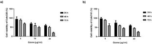
This study aimed to investigate the effects of ozone exposure on apoptotic mechanisms in primary (Colo-320) and metastatic (Colo-741) cell lines. Colo-320 and Colo-741 cells were grown in RPMI-1640 medium supplemented with 10% fetal bovine serum and treated with 20 µg/mL ozone for 72 h after MTT assay. Inverted microscopy was used for morphological assessment, and immunocytochemistry followed by H-SCORE analysis was performed to measure the expression of apoptotic markers. The statistical analysis was conducted using unpaired T-tests or Mann–Whitney U tests, with significance set at p < 0.05. Morphological analysis showed no significant changes in cell shape in Colo-320 or Colo-741 cells after ozone exposure. However, immunocytochemical analysis revealed significant increases in semi-quantitative histological scores (H-SCORES) for cleaved caspase-3, Bax, and cytochrome c in both cell lines, indicating enhanced apoptotic activity (p < 0.05). Conversely, Bcl-2 expression was significantly decreased in both Colo-320 and Colo-741 cell lines after ozone exposure (p < 0.05). Ozone exposure promoted apoptosis in both Colo-320 and Colo-741 cell lines, as evidenced by increased pro-apoptotic markers and decreased anti-apoptotic markers. These molecular changes were notable, yet they did not visibly alter cell morphology. The observed similarities between Colo-320 and Colo-741 cell responses suggest the need for further investigation. These findings indicate that ozone exposure may influence tumor cell apoptosis while preserving cell structure, highlighting the necessity of further research into its potential therapeutic implications.
期刊介绍:
The Journal of Molecular Histology publishes results of original research on the localization and expression of molecules in animal cells, tissues and organs. Coverage includes studies describing novel cellular or ultrastructural distributions of molecules which provide insight into biochemical or physiological function, development, histologic structure and disease processes.
Major research themes of particular interest include:
- Cell-Cell and Cell-Matrix Interactions;
- Connective Tissues;
- Development and Disease;
- Neuroscience.
Please note that the Journal of Molecular Histology does not consider manuscripts dealing with the application of immunological or other probes on non-standard laboratory animal models unless the results are clearly of significant and general biological importance.
The Journal of Molecular Histology publishes full-length original research papers, review articles, short communications and letters to the editors. All manuscripts are typically reviewed by two independent referees. The Journal of Molecular Histology is a continuation of The Histochemical Journal.

 求助内容:
求助内容: 应助结果提醒方式:


