Krisztina Mikulás, Xinyi Qian, Péter Tajti, Gergely Agócs, German O. Gallucci, Ignacio Pedrinaci, Péter Hermann
{"title":"用于捕获种植体周围涌现轮廓的数字印象方法的准确性:系统综述","authors":"Krisztina Mikulás, Xinyi Qian, Péter Tajti, Gergely Agócs, German O. Gallucci, Ignacio Pedrinaci, Péter Hermann","doi":"10.1111/clr.14441","DOIUrl":null,"url":null,"abstract":"<div>\n \n \n <section>\n \n <h3> Background</h3>\n \n <p>Accurately replicating the emergence profile (EP) of conditioned soft tissue is pivotal for the success of implant-supported restorations. In the field of digital technology, various methods have emerged to capture EP. This review aims to critically assess current digital methodologies for capturing peri-implant EP.</p>\n </section>\n \n <section>\n \n <h3> Material and Methods</h3>\n \n <p>Prospective interventional or observational clinical studies focusing on digitally mapping the emergence profile (EP) around single implant-supported restorations were included. Systematic reviews, in vitro and animal studies, and those not emphasizing EP capture were excluded. A systematic search across four databases (MEDLINE, CENTRAL, Embase, Web of Science) was conducted on 7th August 2024 based on a previously registered protocol (PROSPERO registration number: CRD42023459484). Risk of bias was assessed with RoB 2, ROBINS-I, and JBI critical appraisal tools. Qualitative and quantitative analyses were carried out.</p>\n </section>\n \n <section>\n \n <h3> Results</h3>\n \n <p>Twenty-four eligible studies were identified, comprising 5 dental techniques, 12 case reports, 1 randomized controlled study, 2 cross-sectional studies, and 4 cross-over studies. The studies reported semi-digital pathways, direct scanning, indirect scanning, coded-healing abutments, and individualized use of scan bodies. Notably, the direct scanning technique showed considerable soft tissue collapse. Similar results can be achieved with indirect scanning and the conventional method.</p>\n </section>\n \n <section>\n \n <h3> Conclusions</h3>\n \n <p>Indirect EP scanning appears as the most promising method for capturing peri-implant EP. However, a confirmation of this finding requires a quantitative analysis through randomized clinical trials.</p>\n </section>\n </div>","PeriodicalId":10455,"journal":{"name":"Clinical Oral Implants Research","volume":"36 8","pages":"930-943"},"PeriodicalIF":5.3000,"publicationDate":"2025-04-12","publicationTypes":"Journal Article","fieldsOfStudy":null,"isOpenAccess":false,"openAccessPdf":"https://onlinelibrary.wiley.com/doi/epdf/10.1111/clr.14441","citationCount":"0","resultStr":"{\"title\":\"Accuracy of Digital Impression Methods for Capturing the Peri-Implant Emergence Profile: A Systematic Review\",\"authors\":\"Krisztina Mikulás, Xinyi Qian, Péter Tajti, Gergely Agócs, German O. Gallucci, Ignacio Pedrinaci, Péter Hermann\",\"doi\":\"10.1111/clr.14441\",\"DOIUrl\":null,\"url\":null,\"abstract\":\"<div>\\n \\n \\n <section>\\n \\n <h3> Background</h3>\\n \\n <p>Accurately replicating the emergence profile (EP) of conditioned soft tissue is pivotal for the success of implant-supported restorations. In the field of digital technology, various methods have emerged to capture EP. This review aims to critically assess current digital methodologies for capturing peri-implant EP.</p>\\n </section>\\n \\n <section>\\n \\n <h3> Material and Methods</h3>\\n \\n <p>Prospective interventional or observational clinical studies focusing on digitally mapping the emergence profile (EP) around single implant-supported restorations were included. Systematic reviews, in vitro and animal studies, and those not emphasizing EP capture were excluded. A systematic search across four databases (MEDLINE, CENTRAL, Embase, Web of Science) was conducted on 7th August 2024 based on a previously registered protocol (PROSPERO registration number: CRD42023459484). Risk of bias was assessed with RoB 2, ROBINS-I, and JBI critical appraisal tools. Qualitative and quantitative analyses were carried out.</p>\\n </section>\\n \\n <section>\\n \\n <h3> Results</h3>\\n \\n <p>Twenty-four eligible studies were identified, comprising 5 dental techniques, 12 case reports, 1 randomized controlled study, 2 cross-sectional studies, and 4 cross-over studies. The studies reported semi-digital pathways, direct scanning, indirect scanning, coded-healing abutments, and individualized use of scan bodies. Notably, the direct scanning technique showed considerable soft tissue collapse. Similar results can be achieved with indirect scanning and the conventional method.</p>\\n </section>\\n \\n <section>\\n \\n <h3> Conclusions</h3>\\n \\n <p>Indirect EP scanning appears as the most promising method for capturing peri-implant EP. However, a confirmation of this finding requires a quantitative analysis through randomized clinical trials.</p>\\n </section>\\n </div>\",\"PeriodicalId\":10455,\"journal\":{\"name\":\"Clinical Oral Implants Research\",\"volume\":\"36 8\",\"pages\":\"930-943\"},\"PeriodicalIF\":5.3000,\"publicationDate\":\"2025-04-12\",\"publicationTypes\":\"Journal Article\",\"fieldsOfStudy\":null,\"isOpenAccess\":false,\"openAccessPdf\":\"https://onlinelibrary.wiley.com/doi/epdf/10.1111/clr.14441\",\"citationCount\":\"0\",\"resultStr\":null,\"platform\":\"Semanticscholar\",\"paperid\":null,\"PeriodicalName\":\"Clinical Oral Implants Research\",\"FirstCategoryId\":\"5\",\"ListUrlMain\":\"https://onlinelibrary.wiley.com/doi/10.1111/clr.14441\",\"RegionNum\":1,\"RegionCategory\":\"医学\",\"ArticlePicture\":[],\"TitleCN\":null,\"AbstractTextCN\":null,\"PMCID\":null,\"EPubDate\":\"\",\"PubModel\":\"\",\"JCR\":\"Q1\",\"JCRName\":\"DENTISTRY, ORAL SURGERY & MEDICINE\",\"Score\":null,\"Total\":0}","platform":"Semanticscholar","paperid":null,"PeriodicalName":"Clinical Oral Implants Research","FirstCategoryId":"5","ListUrlMain":"https://onlinelibrary.wiley.com/doi/10.1111/clr.14441","RegionNum":1,"RegionCategory":"医学","ArticlePicture":[],"TitleCN":null,"AbstractTextCN":null,"PMCID":null,"EPubDate":"","PubModel":"","JCR":"Q1","JCRName":"DENTISTRY, ORAL SURGERY & MEDICINE","Score":null,"Total":0}
引用次数: 0
摘要
准确复制条件软组织的涌现特征(EP)对于种植体支持修复的成功至关重要。在数字技术领域,出现了各种捕获EP的方法。本综述旨在批判性地评估当前用于捕获植入期EP的数字方法。材料和方法前瞻性干预性或观察性临床研究聚焦于单种植体支撑修复体周围涌现剖面(EP)的数字测绘。排除了系统评价、体外和动物研究以及不强调EP捕获的研究。基于先前注册的协议(PROSPERO注册号:CRD42023459484),于2024年8月7日对四个数据库(MEDLINE, CENTRAL, Embase, Web of Science)进行了系统搜索。采用rob2、ROBINS‐I和JBI关键评估工具评估偏倚风险。进行了定性和定量分析。结果共纳入24项符合条件的研究,包括5项牙科技术、12例病例报告、1项随机对照研究、2项横断面研究和4项交叉研究。这些研究报告了半数字通路、直接扫描、间接扫描、编码愈合基台和扫描体的个性化使用。值得注意的是,直接扫描技术显示相当大的软组织塌陷。间接扫描和常规扫描均可获得相似的结果。结论直接电位扫描是一种最有前途的捕获种植体周围电位的方法。然而,要证实这一发现,需要通过随机临床试验进行定量分析。
Accuracy of Digital Impression Methods for Capturing the Peri-Implant Emergence Profile: A Systematic Review
Background
Accurately replicating the emergence profile (EP) of conditioned soft tissue is pivotal for the success of implant-supported restorations. In the field of digital technology, various methods have emerged to capture EP. This review aims to critically assess current digital methodologies for capturing peri-implant EP.
Material and Methods
Prospective interventional or observational clinical studies focusing on digitally mapping the emergence profile (EP) around single implant-supported restorations were included. Systematic reviews, in vitro and animal studies, and those not emphasizing EP capture were excluded. A systematic search across four databases (MEDLINE, CENTRAL, Embase, Web of Science) was conducted on 7th August 2024 based on a previously registered protocol (PROSPERO registration number: CRD42023459484). Risk of bias was assessed with RoB 2, ROBINS-I, and JBI critical appraisal tools. Qualitative and quantitative analyses were carried out.
Results
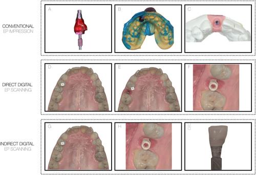
Twenty-four eligible studies were identified, comprising 5 dental techniques, 12 case reports, 1 randomized controlled study, 2 cross-sectional studies, and 4 cross-over studies. The studies reported semi-digital pathways, direct scanning, indirect scanning, coded-healing abutments, and individualized use of scan bodies. Notably, the direct scanning technique showed considerable soft tissue collapse. Similar results can be achieved with indirect scanning and the conventional method.
Conclusions
Indirect EP scanning appears as the most promising method for capturing peri-implant EP. However, a confirmation of this finding requires a quantitative analysis through randomized clinical trials.
期刊介绍:
Clinical Oral Implants Research conveys scientific progress in the field of implant dentistry and its related areas to clinicians, teachers and researchers concerned with the application of this information for the benefit of patients in need of oral implants. The journal addresses itself to clinicians, general practitioners, periodontists, oral and maxillofacial surgeons and prosthodontists, as well as to teachers, academicians and scholars involved in the education of professionals and in the scientific promotion of the field of implant dentistry.

 求助内容:
求助内容: 应助结果提醒方式:


