{"title":"Hounsfield 单位可预测辅助照射后胃癌幸存者的椎体压缩性骨折。","authors":"Pervin Hurmuz, Yasin Ozyurek, Ecem Yigit, Suayib Yalcin, Fazli Yagiz Yedekci, Faruk Zorlu, Mustafa Cengiz","doi":"10.3857/roj.2024.00409","DOIUrl":null,"url":null,"abstract":"<p><strong>Purpose: </strong>This study aimed to investigate the risk factors and predictive value of vertebral Hounsfield units (HUs) for vertebral compression fracture (VCF) development in gastric cancer (GC) patients who received adjuvant radiotherapy (RT).</p><p><strong>Materials and methods: </strong>We retrospectively analyzed the data of 271 patients with non-metastatic GC who received adjuvant RT between 2010 and 2020. The vertebral bodies from 9th thoracic (T9) to 2nd lumbar (L2) were contoured in computed tomographies used for RT planning, and V30, V35, V40, mean doses, and HUs of vertebrae were documented. We conducted univariate and multivariate analyses to identify the risk factors for VCF development.</p><p><strong>Results: </strong>The median follow-up time was 35.7 months. VCF developed in 23 patients (8.5%) in a median of 30.6 months (range, 3.4 to 117.3) after the end of RT. In total, 37 vertebrae were fractured, with 14 located in T12, nine in L1, seven in T11, four in L2, and three in T10. Older age, female sex, non-smoking status, and lower median vertebrae HUs were significantly associated with VCF in the univariate analysis. In the multivariate analysis, lower median HUs of T12 vertebrae (odds ratio, 0.965; 95% confidence interval, 0.942 to 0.989; p = 0.004) remained significant. The optimal cut-off value for T12 HU was 205.1, with an area under the receiver operating characteristic curve of 0.765, sensitivity of 85.7%, and specificity of 65%.</p><p><strong>Conclusion: </strong>The lower median HU value of T12 vertebrae is a significant and independent risk factor for VCF development in GC patients who received adjuvant RT. HUs values serve as a simple and reliable predictor of VCF development in this population.</p>","PeriodicalId":94184,"journal":{"name":"Radiation oncology journal","volume":"43 1","pages":"30-39"},"PeriodicalIF":0.0000,"publicationDate":"2025-03-01","publicationTypes":"Journal Article","fieldsOfStudy":null,"isOpenAccess":false,"openAccessPdf":"https://www.ncbi.nlm.nih.gov/pmc/articles/PMC12010886/pdf/","citationCount":"0","resultStr":"{\"title\":\"Hounsfield units predict vertebral compression fractures in gastric cancer survivors after adjuvant irradiation.\",\"authors\":\"Pervin Hurmuz, Yasin Ozyurek, Ecem Yigit, Suayib Yalcin, Fazli Yagiz Yedekci, Faruk Zorlu, Mustafa Cengiz\",\"doi\":\"10.3857/roj.2024.00409\",\"DOIUrl\":null,\"url\":null,\"abstract\":\"<p><strong>Purpose: </strong>This study aimed to investigate the risk factors and predictive value of vertebral Hounsfield units (HUs) for vertebral compression fracture (VCF) development in gastric cancer (GC) patients who received adjuvant radiotherapy (RT).</p><p><strong>Materials and methods: </strong>We retrospectively analyzed the data of 271 patients with non-metastatic GC who received adjuvant RT between 2010 and 2020. The vertebral bodies from 9th thoracic (T9) to 2nd lumbar (L2) were contoured in computed tomographies used for RT planning, and V30, V35, V40, mean doses, and HUs of vertebrae were documented. We conducted univariate and multivariate analyses to identify the risk factors for VCF development.</p><p><strong>Results: </strong>The median follow-up time was 35.7 months. VCF developed in 23 patients (8.5%) in a median of 30.6 months (range, 3.4 to 117.3) after the end of RT. In total, 37 vertebrae were fractured, with 14 located in T12, nine in L1, seven in T11, four in L2, and three in T10. Older age, female sex, non-smoking status, and lower median vertebrae HUs were significantly associated with VCF in the univariate analysis. In the multivariate analysis, lower median HUs of T12 vertebrae (odds ratio, 0.965; 95% confidence interval, 0.942 to 0.989; p = 0.004) remained significant. The optimal cut-off value for T12 HU was 205.1, with an area under the receiver operating characteristic curve of 0.765, sensitivity of 85.7%, and specificity of 65%.</p><p><strong>Conclusion: </strong>The lower median HU value of T12 vertebrae is a significant and independent risk factor for VCF development in GC patients who received adjuvant RT. HUs values serve as a simple and reliable predictor of VCF development in this population.</p>\",\"PeriodicalId\":94184,\"journal\":{\"name\":\"Radiation oncology journal\",\"volume\":\"43 1\",\"pages\":\"30-39\"},\"PeriodicalIF\":0.0000,\"publicationDate\":\"2025-03-01\",\"publicationTypes\":\"Journal Article\",\"fieldsOfStudy\":null,\"isOpenAccess\":false,\"openAccessPdf\":\"https://www.ncbi.nlm.nih.gov/pmc/articles/PMC12010886/pdf/\",\"citationCount\":\"0\",\"resultStr\":null,\"platform\":\"Semanticscholar\",\"paperid\":null,\"PeriodicalName\":\"Radiation oncology journal\",\"FirstCategoryId\":\"1085\",\"ListUrlMain\":\"https://doi.org/10.3857/roj.2024.00409\",\"RegionNum\":0,\"RegionCategory\":null,\"ArticlePicture\":[],\"TitleCN\":null,\"AbstractTextCN\":null,\"PMCID\":null,\"EPubDate\":\"2025/2/26 0:00:00\",\"PubModel\":\"Epub\",\"JCR\":\"\",\"JCRName\":\"\",\"Score\":null,\"Total\":0}","platform":"Semanticscholar","paperid":null,"PeriodicalName":"Radiation oncology journal","FirstCategoryId":"1085","ListUrlMain":"https://doi.org/10.3857/roj.2024.00409","RegionNum":0,"RegionCategory":null,"ArticlePicture":[],"TitleCN":null,"AbstractTextCN":null,"PMCID":null,"EPubDate":"2025/2/26 0:00:00","PubModel":"Epub","JCR":"","JCRName":"","Score":null,"Total":0}
Hounsfield units predict vertebral compression fractures in gastric cancer survivors after adjuvant irradiation.
Purpose: This study aimed to investigate the risk factors and predictive value of vertebral Hounsfield units (HUs) for vertebral compression fracture (VCF) development in gastric cancer (GC) patients who received adjuvant radiotherapy (RT).
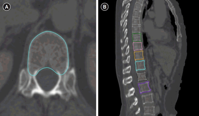
Materials and methods: We retrospectively analyzed the data of 271 patients with non-metastatic GC who received adjuvant RT between 2010 and 2020. The vertebral bodies from 9th thoracic (T9) to 2nd lumbar (L2) were contoured in computed tomographies used for RT planning, and V30, V35, V40, mean doses, and HUs of vertebrae were documented. We conducted univariate and multivariate analyses to identify the risk factors for VCF development.
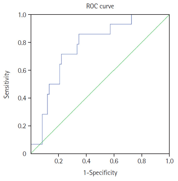
Results: The median follow-up time was 35.7 months. VCF developed in 23 patients (8.5%) in a median of 30.6 months (range, 3.4 to 117.3) after the end of RT. In total, 37 vertebrae were fractured, with 14 located in T12, nine in L1, seven in T11, four in L2, and three in T10. Older age, female sex, non-smoking status, and lower median vertebrae HUs were significantly associated with VCF in the univariate analysis. In the multivariate analysis, lower median HUs of T12 vertebrae (odds ratio, 0.965; 95% confidence interval, 0.942 to 0.989; p = 0.004) remained significant. The optimal cut-off value for T12 HU was 205.1, with an area under the receiver operating characteristic curve of 0.765, sensitivity of 85.7%, and specificity of 65%.
Conclusion: The lower median HU value of T12 vertebrae is a significant and independent risk factor for VCF development in GC patients who received adjuvant RT. HUs values serve as a simple and reliable predictor of VCF development in this population.

 求助内容:
求助内容: 应助结果提醒方式:


