Pavel Pavlov, Andreas Kontny, Neele Wagner, Nikola Kolev, Alexander Zlatarov, Turgay Kalinov, Anton B Tonchev
{"title":"使用改进的基于立方体的组织清除技术的人类结肠组织的三维可视化。","authors":"Pavel Pavlov, Andreas Kontny, Neele Wagner, Nikola Kolev, Alexander Zlatarov, Turgay Kalinov, Anton B Tonchev","doi":"10.14440/jbm.2025.0101","DOIUrl":null,"url":null,"abstract":"<p><strong>Background: </strong>Colorectal cancer represents one of the most common neoplastic diseases worldwide, making it a frequent focus in routine pathological analyses. Visualizing complex three-dimensional (3D) structures, such as nerves within tumors, requires thick tissue sections, which necessitates the use of optical tissue-clearing methods to achieve transparency. However, following tissue clearing, samples typically require advanced imaging techniques such as light-sheet and two-photon confocal microscopy, which are usually unavailable in standard histological laboratories.</p><p><strong>Objective: </strong>We aimed to demonstrate how a well-established tissue-clearing approach can be adapted for use in a routine histological laboratory, enabling a robust 3D visualization of nerve fibers in samples of both normal human colon and colon cancer tissues.</p><p><strong>Methods: </strong>We modified the \"clear unobstructed brain/body imaging cocktails\" method, originally developed for whole-brain imaging in mice, and applied it to human colon tissue samples measuring approximately 10 mm<sup>3</sup>, a standard size typically processed in pathological laboratories.</p><p><strong>Results: </strong>Our protocol, which integrates a tissue-clearing technique, enabled reliable immunofluorescent visualization of colonic nerve fibers labeled with anti-β<sub>3</sub>-tubulin antibodies. The labeled nerve fibers could be observed using a standard epifluorescence microscope, and high-quality 3D reconstructions were generated through a simple image analysis approach using the open-source software ilastik, which eliminates the need for confocal microscopy.</p><p><strong>Conclusion: </strong>The proposed steps provide a valuable method for researchers to visualize complex 3D structures, such as neural cells and processes, in both normal and tumor-transformed tissue settings.</p>","PeriodicalId":73618,"journal":{"name":"Journal of biological methods","volume":"12 1","pages":"e99010052"},"PeriodicalIF":0.0000,"publicationDate":"2025-02-04","publicationTypes":"Journal Article","fieldsOfStudy":null,"isOpenAccess":false,"openAccessPdf":"https://www.ncbi.nlm.nih.gov/pmc/articles/PMC11973050/pdf/","citationCount":"0","resultStr":"{\"title\":\"3D visualization of human colon tissue using a modified CUBIC-based tissue-clearing technique.\",\"authors\":\"Pavel Pavlov, Andreas Kontny, Neele Wagner, Nikola Kolev, Alexander Zlatarov, Turgay Kalinov, Anton B Tonchev\",\"doi\":\"10.14440/jbm.2025.0101\",\"DOIUrl\":null,\"url\":null,\"abstract\":\"<p><strong>Background: </strong>Colorectal cancer represents one of the most common neoplastic diseases worldwide, making it a frequent focus in routine pathological analyses. Visualizing complex three-dimensional (3D) structures, such as nerves within tumors, requires thick tissue sections, which necessitates the use of optical tissue-clearing methods to achieve transparency. However, following tissue clearing, samples typically require advanced imaging techniques such as light-sheet and two-photon confocal microscopy, which are usually unavailable in standard histological laboratories.</p><p><strong>Objective: </strong>We aimed to demonstrate how a well-established tissue-clearing approach can be adapted for use in a routine histological laboratory, enabling a robust 3D visualization of nerve fibers in samples of both normal human colon and colon cancer tissues.</p><p><strong>Methods: </strong>We modified the \\\"clear unobstructed brain/body imaging cocktails\\\" method, originally developed for whole-brain imaging in mice, and applied it to human colon tissue samples measuring approximately 10 mm<sup>3</sup>, a standard size typically processed in pathological laboratories.</p><p><strong>Results: </strong>Our protocol, which integrates a tissue-clearing technique, enabled reliable immunofluorescent visualization of colonic nerve fibers labeled with anti-β<sub>3</sub>-tubulin antibodies. The labeled nerve fibers could be observed using a standard epifluorescence microscope, and high-quality 3D reconstructions were generated through a simple image analysis approach using the open-source software ilastik, which eliminates the need for confocal microscopy.</p><p><strong>Conclusion: </strong>The proposed steps provide a valuable method for researchers to visualize complex 3D structures, such as neural cells and processes, in both normal and tumor-transformed tissue settings.</p>\",\"PeriodicalId\":73618,\"journal\":{\"name\":\"Journal of biological methods\",\"volume\":\"12 1\",\"pages\":\"e99010052\"},\"PeriodicalIF\":0.0000,\"publicationDate\":\"2025-02-04\",\"publicationTypes\":\"Journal Article\",\"fieldsOfStudy\":null,\"isOpenAccess\":false,\"openAccessPdf\":\"https://www.ncbi.nlm.nih.gov/pmc/articles/PMC11973050/pdf/\",\"citationCount\":\"0\",\"resultStr\":null,\"platform\":\"Semanticscholar\",\"paperid\":null,\"PeriodicalName\":\"Journal of biological methods\",\"FirstCategoryId\":\"1085\",\"ListUrlMain\":\"https://doi.org/10.14440/jbm.2025.0101\",\"RegionNum\":0,\"RegionCategory\":null,\"ArticlePicture\":[],\"TitleCN\":null,\"AbstractTextCN\":null,\"PMCID\":null,\"EPubDate\":\"2025/1/1 0:00:00\",\"PubModel\":\"eCollection\",\"JCR\":\"\",\"JCRName\":\"\",\"Score\":null,\"Total\":0}","platform":"Semanticscholar","paperid":null,"PeriodicalName":"Journal of biological methods","FirstCategoryId":"1085","ListUrlMain":"https://doi.org/10.14440/jbm.2025.0101","RegionNum":0,"RegionCategory":null,"ArticlePicture":[],"TitleCN":null,"AbstractTextCN":null,"PMCID":null,"EPubDate":"2025/1/1 0:00:00","PubModel":"eCollection","JCR":"","JCRName":"","Score":null,"Total":0}
3D visualization of human colon tissue using a modified CUBIC-based tissue-clearing technique.
Background: Colorectal cancer represents one of the most common neoplastic diseases worldwide, making it a frequent focus in routine pathological analyses. Visualizing complex three-dimensional (3D) structures, such as nerves within tumors, requires thick tissue sections, which necessitates the use of optical tissue-clearing methods to achieve transparency. However, following tissue clearing, samples typically require advanced imaging techniques such as light-sheet and two-photon confocal microscopy, which are usually unavailable in standard histological laboratories.
Objective: We aimed to demonstrate how a well-established tissue-clearing approach can be adapted for use in a routine histological laboratory, enabling a robust 3D visualization of nerve fibers in samples of both normal human colon and colon cancer tissues.
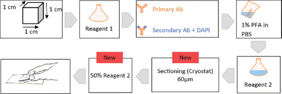
Methods: We modified the "clear unobstructed brain/body imaging cocktails" method, originally developed for whole-brain imaging in mice, and applied it to human colon tissue samples measuring approximately 10 mm3, a standard size typically processed in pathological laboratories.
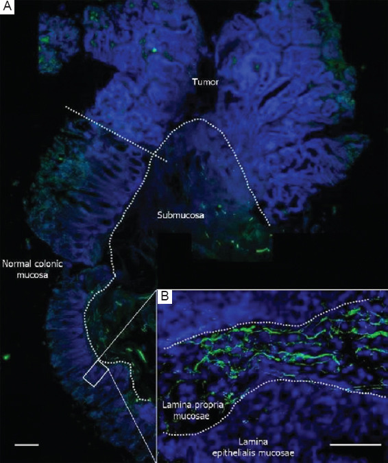
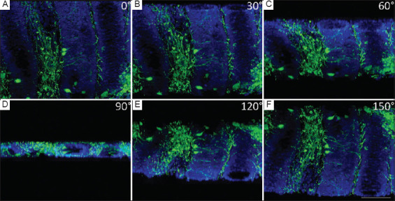
Results: Our protocol, which integrates a tissue-clearing technique, enabled reliable immunofluorescent visualization of colonic nerve fibers labeled with anti-β3-tubulin antibodies. The labeled nerve fibers could be observed using a standard epifluorescence microscope, and high-quality 3D reconstructions were generated through a simple image analysis approach using the open-source software ilastik, which eliminates the need for confocal microscopy.
Conclusion: The proposed steps provide a valuable method for researchers to visualize complex 3D structures, such as neural cells and processes, in both normal and tumor-transformed tissue settings.

 求助内容:
求助内容: 应助结果提醒方式:


