Théo Richard, Victor de Villedon de Naide, Victor Nogues, Thaïs Génisson, Kalvin Narceau, Kun He, Rabea Klaar, Baptiste Durand, Thibault Boullé, Guillaume Poirot, Soumaya Sridi, Jean-David Maes, Marion Constantin, Kinan Kneizeh, Konstantinos Vlachos, Guido Caluori, Pierre Jaïs, Matthias Stuber, Hubert Cochet, Aurelien Bustin
{"title":"利用关节亮血和黑血晚期钆增强MRI改进和自动检测乳头状肌梗死。","authors":"Théo Richard, Victor de Villedon de Naide, Victor Nogues, Thaïs Génisson, Kalvin Narceau, Kun He, Rabea Klaar, Baptiste Durand, Thibault Boullé, Guillaume Poirot, Soumaya Sridi, Jean-David Maes, Marion Constantin, Kinan Kneizeh, Konstantinos Vlachos, Guido Caluori, Pierre Jaïs, Matthias Stuber, Hubert Cochet, Aurelien Bustin","doi":"10.1002/jmri.29777","DOIUrl":null,"url":null,"abstract":"<p><strong>Background: </strong>Papillary muscle infarction (PMI) has been linked to significantly increased mortality and is associated with ventricular arrhythmias and mitral regurgitation. Reference bright-blood late gadolinium enhancement (LGE) imaging provides poor scar-to-blood contrast, making PMI visualization challenging. Black-blood LGE imaging overcomes this limitation by improving the blood-scar contrast.</p><p><strong>Purpose: </strong>To evaluate a recent co-registered bright- (papillary muscle localization) and black-blood (PMI visualization) sequence (Scar-specific imaging with Preserved myOcardial visualizaTion: SPOT) to improve PMI visualization compared to a reference standard phase-sensitive inversion recovery (PSIR) sequence, and to enable automated PMI detection (auto-PMI).</p><p><strong>Study type: </strong>Retrospective.</p><p><strong>Population: </strong>198 patients with ischemic heart disease were divided into an optimization dataset (N = 127) and a testing dataset (N = 71).</p><p><strong>Field strength/sequence: </strong>2D SPOT and PSIR balanced steady-state free precession sequences at 1.5 T.</p><p><strong>Assessment: </strong>Auto-PMI included: image acquisition, slice selection, endocardial segmentation, blood pool preprocessing, and PMI detection. Three radiologists (8, 5 and 2 years of MRI experience) assessed PMI in SPOT and PSIR images independently. A consensus reading regarding all assessments of both sequences was established. The number of patients with PMI in SPOT and PSIR acquisitions was compared. The diagnostic performances of visual (SPOT and PSIR) and auto-PMI (SPOT) detection were evaluated. Inter- and intra-observer reproducibility of the visual PMI detection was assessed.</p><p><strong>Statistical tests: </strong>McNemar test, p-value < 0.05 was considered statistically significant.</p><p><strong>Results: </strong>In the testing dataset, significantly more patients with PMI were detected using SPOT compared to PSIR in each session (37 vs. 27, 36 vs. 29, 41 vs. 31, 42 vs. 25). Sensitivity ranges for visual PMI detection were significantly higher using SPOT (89%-100% vs. 61%-82%). SPOT vs. PSIR inter- and intra-observer reproducibility ranges were 77%-80% vs. 71%-77%, and 97% vs. 88%, respectively. Auto-PMI sensitivity was 87%.</p><p><strong>Data conclusion: </strong>Co-registered bright- and black-blood SPOT imaging improved visual PMI detection and facilitated automated PMI assessment.</p><p><strong>Evidence level: </strong>3. Technical Efficacy: Stage 2.</p>","PeriodicalId":16140,"journal":{"name":"Journal of Magnetic Resonance Imaging","volume":" ","pages":"827-839"},"PeriodicalIF":3.5000,"publicationDate":"2025-09-01","publicationTypes":"Journal Article","fieldsOfStudy":null,"isOpenAccess":false,"openAccessPdf":"https://www.ncbi.nlm.nih.gov/pmc/articles/PMC12335337/pdf/","citationCount":"0","resultStr":"{\"title\":\"Improved and Automated Detection of Papillary Muscle Infarction Using Joint Bright- and Black-Blood Late Gadolinium Enhancement MRI.\",\"authors\":\"Théo Richard, Victor de Villedon de Naide, Victor Nogues, Thaïs Génisson, Kalvin Narceau, Kun He, Rabea Klaar, Baptiste Durand, Thibault Boullé, Guillaume Poirot, Soumaya Sridi, Jean-David Maes, Marion Constantin, Kinan Kneizeh, Konstantinos Vlachos, Guido Caluori, Pierre Jaïs, Matthias Stuber, Hubert Cochet, Aurelien Bustin\",\"doi\":\"10.1002/jmri.29777\",\"DOIUrl\":null,\"url\":null,\"abstract\":\"<p><strong>Background: </strong>Papillary muscle infarction (PMI) has been linked to significantly increased mortality and is associated with ventricular arrhythmias and mitral regurgitation. Reference bright-blood late gadolinium enhancement (LGE) imaging provides poor scar-to-blood contrast, making PMI visualization challenging. Black-blood LGE imaging overcomes this limitation by improving the blood-scar contrast.</p><p><strong>Purpose: </strong>To evaluate a recent co-registered bright- (papillary muscle localization) and black-blood (PMI visualization) sequence (Scar-specific imaging with Preserved myOcardial visualizaTion: SPOT) to improve PMI visualization compared to a reference standard phase-sensitive inversion recovery (PSIR) sequence, and to enable automated PMI detection (auto-PMI).</p><p><strong>Study type: </strong>Retrospective.</p><p><strong>Population: </strong>198 patients with ischemic heart disease were divided into an optimization dataset (N = 127) and a testing dataset (N = 71).</p><p><strong>Field strength/sequence: </strong>2D SPOT and PSIR balanced steady-state free precession sequences at 1.5 T.</p><p><strong>Assessment: </strong>Auto-PMI included: image acquisition, slice selection, endocardial segmentation, blood pool preprocessing, and PMI detection. Three radiologists (8, 5 and 2 years of MRI experience) assessed PMI in SPOT and PSIR images independently. A consensus reading regarding all assessments of both sequences was established. The number of patients with PMI in SPOT and PSIR acquisitions was compared. The diagnostic performances of visual (SPOT and PSIR) and auto-PMI (SPOT) detection were evaluated. Inter- and intra-observer reproducibility of the visual PMI detection was assessed.</p><p><strong>Statistical tests: </strong>McNemar test, p-value < 0.05 was considered statistically significant.</p><p><strong>Results: </strong>In the testing dataset, significantly more patients with PMI were detected using SPOT compared to PSIR in each session (37 vs. 27, 36 vs. 29, 41 vs. 31, 42 vs. 25). Sensitivity ranges for visual PMI detection were significantly higher using SPOT (89%-100% vs. 61%-82%). SPOT vs. PSIR inter- and intra-observer reproducibility ranges were 77%-80% vs. 71%-77%, and 97% vs. 88%, respectively. Auto-PMI sensitivity was 87%.</p><p><strong>Data conclusion: </strong>Co-registered bright- and black-blood SPOT imaging improved visual PMI detection and facilitated automated PMI assessment.</p><p><strong>Evidence level: </strong>3. Technical Efficacy: Stage 2.</p>\",\"PeriodicalId\":16140,\"journal\":{\"name\":\"Journal of Magnetic Resonance Imaging\",\"volume\":\" \",\"pages\":\"827-839\"},\"PeriodicalIF\":3.5000,\"publicationDate\":\"2025-09-01\",\"publicationTypes\":\"Journal Article\",\"fieldsOfStudy\":null,\"isOpenAccess\":false,\"openAccessPdf\":\"https://www.ncbi.nlm.nih.gov/pmc/articles/PMC12335337/pdf/\",\"citationCount\":\"0\",\"resultStr\":null,\"platform\":\"Semanticscholar\",\"paperid\":null,\"PeriodicalName\":\"Journal of Magnetic Resonance Imaging\",\"FirstCategoryId\":\"3\",\"ListUrlMain\":\"https://doi.org/10.1002/jmri.29777\",\"RegionNum\":2,\"RegionCategory\":\"医学\",\"ArticlePicture\":[],\"TitleCN\":null,\"AbstractTextCN\":null,\"PMCID\":null,\"EPubDate\":\"2025/4/9 0:00:00\",\"PubModel\":\"Epub\",\"JCR\":\"Q1\",\"JCRName\":\"RADIOLOGY, NUCLEAR MEDICINE & MEDICAL IMAGING\",\"Score\":null,\"Total\":0}","platform":"Semanticscholar","paperid":null,"PeriodicalName":"Journal of Magnetic Resonance Imaging","FirstCategoryId":"3","ListUrlMain":"https://doi.org/10.1002/jmri.29777","RegionNum":2,"RegionCategory":"医学","ArticlePicture":[],"TitleCN":null,"AbstractTextCN":null,"PMCID":null,"EPubDate":"2025/4/9 0:00:00","PubModel":"Epub","JCR":"Q1","JCRName":"RADIOLOGY, NUCLEAR MEDICINE & MEDICAL IMAGING","Score":null,"Total":0}
引用次数: 0
摘要
背景:乳头状肌梗死(PMI)与死亡率显著增加有关,并与室性心律失常和二尖瓣反流相关。参考亮血晚期钆增强(LGE)成像提供了较差的疤痕-血液对比,使PMI可视化具有挑战性。黑血LGE成像通过改善血-疤痕对比克服了这一限制。目的:评估最近共同注册的亮肌(乳头肌定位)和黑血(PMI可视化)序列(疤痕特异性成像与保留心肌可视化:SPOT),与参考标准相敏反演恢复(PSIR)序列相比,改善PMI可视化,并实现自动PMI检测(auto-PMI)。研究类型:回顾性。人群:198例缺血性心脏病患者分为优化数据集(N = 127)和测试数据集(N = 71)。场强/序列:2D SPOT和PSIR平衡的1.5 T的稳态自由进动序列。评估:自动PMI包括:图像采集、切片选择、心内膜分割、血池预处理和PMI检测。三位放射科医生(分别有8年、5年和2年的MRI经验)独立评估SPOT和PSIR图像中的PMI。建立了关于这两个序列的所有评估的共识读数。比较了SPOT和PSIR采集中PMI患者的数量。评估视觉(SPOT和PSIR)和自动pmi (SPOT)检测的诊断性能。评估了视觉PMI检测在观察者之间和观察者内部的再现性。统计检验:McNemar检验,p值结果:在测试数据集中,与PSIR相比,每次使用SPOT检测到的PMI患者明显更多(37 vs. 27,36 vs. 29,41 vs. 31,42 vs. 25)。使用SPOT进行视觉PMI检测的灵敏度范围明显更高(89%-100% vs 61%-82%)。SPOT和PSIR在观察者之间和观察者内部的重复性范围分别为77%-80%对71%-77%,97%对88%。自动pmi敏感性为87%。数据结论:共同登记的亮血和黑血斑点成像改善了视觉PMI检测,促进了PMI的自动评估。证据等级:3。技术功效:第二阶段。
Improved and Automated Detection of Papillary Muscle Infarction Using Joint Bright- and Black-Blood Late Gadolinium Enhancement MRI.
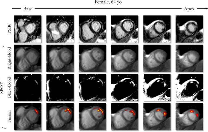
Background: Papillary muscle infarction (PMI) has been linked to significantly increased mortality and is associated with ventricular arrhythmias and mitral regurgitation. Reference bright-blood late gadolinium enhancement (LGE) imaging provides poor scar-to-blood contrast, making PMI visualization challenging. Black-blood LGE imaging overcomes this limitation by improving the blood-scar contrast.
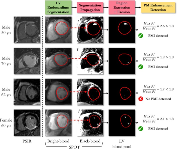
Purpose: To evaluate a recent co-registered bright- (papillary muscle localization) and black-blood (PMI visualization) sequence (Scar-specific imaging with Preserved myOcardial visualizaTion: SPOT) to improve PMI visualization compared to a reference standard phase-sensitive inversion recovery (PSIR) sequence, and to enable automated PMI detection (auto-PMI).
Study type: Retrospective.
Population: 198 patients with ischemic heart disease were divided into an optimization dataset (N = 127) and a testing dataset (N = 71).
Field strength/sequence: 2D SPOT and PSIR balanced steady-state free precession sequences at 1.5 T.
Assessment: Auto-PMI included: image acquisition, slice selection, endocardial segmentation, blood pool preprocessing, and PMI detection. Three radiologists (8, 5 and 2 years of MRI experience) assessed PMI in SPOT and PSIR images independently. A consensus reading regarding all assessments of both sequences was established. The number of patients with PMI in SPOT and PSIR acquisitions was compared. The diagnostic performances of visual (SPOT and PSIR) and auto-PMI (SPOT) detection were evaluated. Inter- and intra-observer reproducibility of the visual PMI detection was assessed.
Statistical tests: McNemar test, p-value < 0.05 was considered statistically significant.
Results: In the testing dataset, significantly more patients with PMI were detected using SPOT compared to PSIR in each session (37 vs. 27, 36 vs. 29, 41 vs. 31, 42 vs. 25). Sensitivity ranges for visual PMI detection were significantly higher using SPOT (89%-100% vs. 61%-82%). SPOT vs. PSIR inter- and intra-observer reproducibility ranges were 77%-80% vs. 71%-77%, and 97% vs. 88%, respectively. Auto-PMI sensitivity was 87%.
Data conclusion: Co-registered bright- and black-blood SPOT imaging improved visual PMI detection and facilitated automated PMI assessment.
期刊介绍:
The Journal of Magnetic Resonance Imaging (JMRI) is an international journal devoted to the timely publication of basic and clinical research, educational and review articles, and other information related to the diagnostic applications of magnetic resonance.

 求助内容:
求助内容: 应助结果提醒方式:


