{"title":"腹腔镜切除脾后硬化性血管瘤样结节转化:病例报告及文献复习。","authors":"Shingo Yamasaki, Hiroto Nishino, Takayuki Anazawa, Yuki Teramoto, Takahiro Nishio, Shoichi Kageyama, Kazuyuki Nagai, Yoichiro Uchida, Hironori Haga, Etsuro Hatano","doi":"10.70352/scrj.cr.24-0057","DOIUrl":null,"url":null,"abstract":"<p><strong>Introduction: </strong>Most splenic tumors are benign; however, it is essential to differentiate them from malignant tumors, such as malignant lymphoma and metastatic tumors. Sclerosing angiomatoid nodular transformation (SANT) is a relatively rare benign tumor that has been reported recently. Splenectomy is performed in most cases of SANT because of the challenges associated with a definitive diagnosis. However, in some cases, SANT can be diagnosed through endoscopic ultrasound-guided fine-needle aspiration (EUS-FNA), and these cases are subsequently followed up. In this report, we present 2 cases of splenic SANT that underwent laparoscopic resection. In Case 1, atypical imaging findings required EUS-FNA for further evaluation. Case 2 exhibited typical imaging findings of SANT, and therefore, EUS-FNA was not performed.</p><p><strong>Case presentation: </strong>Case 1: A 47-year-old female was found to have a 26 mm tumor in the spleen on abdominal ultrasonography during follow-up for gallbladder polyps. Abdominal computed tomography (CT), magnetic resonance imaging (MRI), and positron emission tomography-CT were performed. EUS-FNA was performed because of the high surgical risk associated with pulmonary hypertension and because hemangioendothelioma, an intermediate malignancy, was suspected. Subsequently, laparoscopic splenectomy was performed, and SANT was diagnosed. Case 2: A 46-year-old female had an incidental detection of a tumor in the spleen on CT. SANT was suspected based on CT and MRI findings. Malignancy could not be completely ruled out owing to the gradual growth of the mass; therefore, the patient was referred to our hospital for surgery. Laparoscopic splenectomy was performed, and SANT was subsequently diagnosed.</p><p><strong>Conclusion: </strong>SANT is a benign tumor that is difficult to diagnose; however, in some cases, it can be diagnosed using EUS-FNA. We report 2 cases of SANT of the spleen that underwent laparoscopic resection. In cases where the diagnosis is confirmed through imaging or histological examination, disease management with follow-up and without surgery is a possible alternative.</p>","PeriodicalId":22096,"journal":{"name":"Surgical Case Reports","volume":"11 1","pages":""},"PeriodicalIF":0.7000,"publicationDate":"2025-01-01","publicationTypes":"Journal Article","fieldsOfStudy":null,"isOpenAccess":false,"openAccessPdf":"https://www.ncbi.nlm.nih.gov/pmc/articles/PMC11973248/pdf/","citationCount":"0","resultStr":"{\"title\":\"Sclerosing Angiomatoid Nodular Transformation of the Laparoscopically Resected Spleen: Case Reports and Review of the Literature.\",\"authors\":\"Shingo Yamasaki, Hiroto Nishino, Takayuki Anazawa, Yuki Teramoto, Takahiro Nishio, Shoichi Kageyama, Kazuyuki Nagai, Yoichiro Uchida, Hironori Haga, Etsuro Hatano\",\"doi\":\"10.70352/scrj.cr.24-0057\",\"DOIUrl\":null,\"url\":null,\"abstract\":\"<p><strong>Introduction: </strong>Most splenic tumors are benign; however, it is essential to differentiate them from malignant tumors, such as malignant lymphoma and metastatic tumors. Sclerosing angiomatoid nodular transformation (SANT) is a relatively rare benign tumor that has been reported recently. Splenectomy is performed in most cases of SANT because of the challenges associated with a definitive diagnosis. However, in some cases, SANT can be diagnosed through endoscopic ultrasound-guided fine-needle aspiration (EUS-FNA), and these cases are subsequently followed up. In this report, we present 2 cases of splenic SANT that underwent laparoscopic resection. In Case 1, atypical imaging findings required EUS-FNA for further evaluation. Case 2 exhibited typical imaging findings of SANT, and therefore, EUS-FNA was not performed.</p><p><strong>Case presentation: </strong>Case 1: A 47-year-old female was found to have a 26 mm tumor in the spleen on abdominal ultrasonography during follow-up for gallbladder polyps. Abdominal computed tomography (CT), magnetic resonance imaging (MRI), and positron emission tomography-CT were performed. EUS-FNA was performed because of the high surgical risk associated with pulmonary hypertension and because hemangioendothelioma, an intermediate malignancy, was suspected. Subsequently, laparoscopic splenectomy was performed, and SANT was diagnosed. Case 2: A 46-year-old female had an incidental detection of a tumor in the spleen on CT. SANT was suspected based on CT and MRI findings. Malignancy could not be completely ruled out owing to the gradual growth of the mass; therefore, the patient was referred to our hospital for surgery. Laparoscopic splenectomy was performed, and SANT was subsequently diagnosed.</p><p><strong>Conclusion: </strong>SANT is a benign tumor that is difficult to diagnose; however, in some cases, it can be diagnosed using EUS-FNA. We report 2 cases of SANT of the spleen that underwent laparoscopic resection. In cases where the diagnosis is confirmed through imaging or histological examination, disease management with follow-up and without surgery is a possible alternative.</p>\",\"PeriodicalId\":22096,\"journal\":{\"name\":\"Surgical Case Reports\",\"volume\":\"11 1\",\"pages\":\"\"},\"PeriodicalIF\":0.7000,\"publicationDate\":\"2025-01-01\",\"publicationTypes\":\"Journal Article\",\"fieldsOfStudy\":null,\"isOpenAccess\":false,\"openAccessPdf\":\"https://www.ncbi.nlm.nih.gov/pmc/articles/PMC11973248/pdf/\",\"citationCount\":\"0\",\"resultStr\":null,\"platform\":\"Semanticscholar\",\"paperid\":null,\"PeriodicalName\":\"Surgical Case Reports\",\"FirstCategoryId\":\"1085\",\"ListUrlMain\":\"https://doi.org/10.70352/scrj.cr.24-0057\",\"RegionNum\":0,\"RegionCategory\":null,\"ArticlePicture\":[],\"TitleCN\":null,\"AbstractTextCN\":null,\"PMCID\":null,\"EPubDate\":\"2025/4/2 0:00:00\",\"PubModel\":\"Epub\",\"JCR\":\"Q4\",\"JCRName\":\"SURGERY\",\"Score\":null,\"Total\":0}","platform":"Semanticscholar","paperid":null,"PeriodicalName":"Surgical Case Reports","FirstCategoryId":"1085","ListUrlMain":"https://doi.org/10.70352/scrj.cr.24-0057","RegionNum":0,"RegionCategory":null,"ArticlePicture":[],"TitleCN":null,"AbstractTextCN":null,"PMCID":null,"EPubDate":"2025/4/2 0:00:00","PubModel":"Epub","JCR":"Q4","JCRName":"SURGERY","Score":null,"Total":0}
Sclerosing Angiomatoid Nodular Transformation of the Laparoscopically Resected Spleen: Case Reports and Review of the Literature.
Introduction: Most splenic tumors are benign; however, it is essential to differentiate them from malignant tumors, such as malignant lymphoma and metastatic tumors. Sclerosing angiomatoid nodular transformation (SANT) is a relatively rare benign tumor that has been reported recently. Splenectomy is performed in most cases of SANT because of the challenges associated with a definitive diagnosis. However, in some cases, SANT can be diagnosed through endoscopic ultrasound-guided fine-needle aspiration (EUS-FNA), and these cases are subsequently followed up. In this report, we present 2 cases of splenic SANT that underwent laparoscopic resection. In Case 1, atypical imaging findings required EUS-FNA for further evaluation. Case 2 exhibited typical imaging findings of SANT, and therefore, EUS-FNA was not performed.
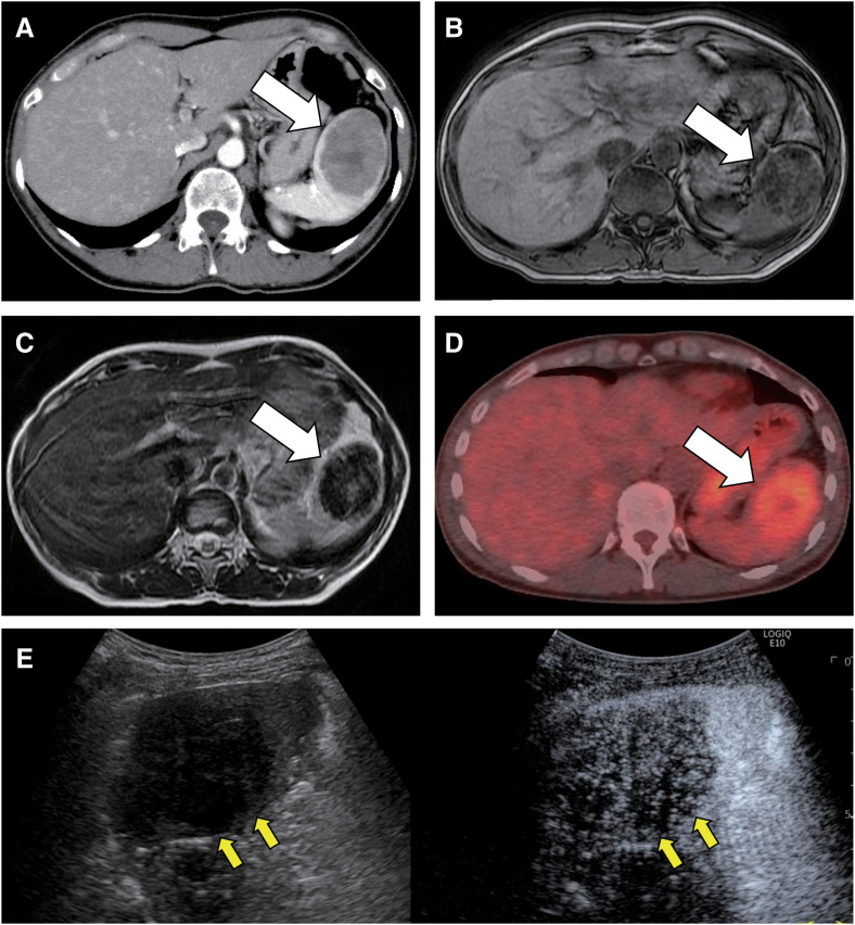
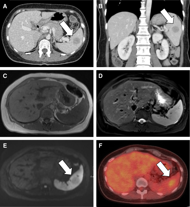
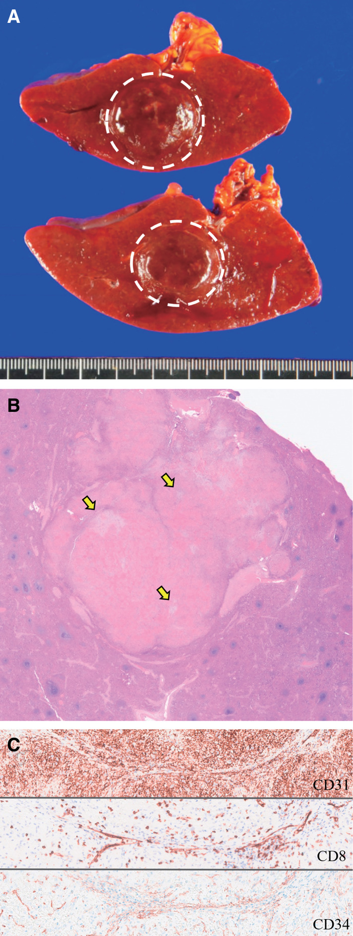
Case presentation: Case 1: A 47-year-old female was found to have a 26 mm tumor in the spleen on abdominal ultrasonography during follow-up for gallbladder polyps. Abdominal computed tomography (CT), magnetic resonance imaging (MRI), and positron emission tomography-CT were performed. EUS-FNA was performed because of the high surgical risk associated with pulmonary hypertension and because hemangioendothelioma, an intermediate malignancy, was suspected. Subsequently, laparoscopic splenectomy was performed, and SANT was diagnosed. Case 2: A 46-year-old female had an incidental detection of a tumor in the spleen on CT. SANT was suspected based on CT and MRI findings. Malignancy could not be completely ruled out owing to the gradual growth of the mass; therefore, the patient was referred to our hospital for surgery. Laparoscopic splenectomy was performed, and SANT was subsequently diagnosed.
Conclusion: SANT is a benign tumor that is difficult to diagnose; however, in some cases, it can be diagnosed using EUS-FNA. We report 2 cases of SANT of the spleen that underwent laparoscopic resection. In cases where the diagnosis is confirmed through imaging or histological examination, disease management with follow-up and without surgery is a possible alternative.

 求助内容:
求助内容: 应助结果提醒方式:


