{"title":"术前磁共振及腹部超声诊断后腹腔镜切除成人回肠囊性重复肠1例。","authors":"Takashi Takeda, Katsuki Danno, Tadafumi Fukata, Itsuko Nakamichi, Kei Yamamoto, Masaya Higashiguchi, Kozo Noguchi, Takafumi Hirao, Mitsugu Sekimoto, Yoshio Oka","doi":"10.70352/scrj.cr.25-0015","DOIUrl":null,"url":null,"abstract":"<p><strong>Introduction: </strong>Small bowel duplication in adults is an uncommon congenital anomaly that often presents with nonspecific symptoms, such as abdominal pain, vomiting, or constipation, which complicates diagnosis. Imaging techniques such as computed tomography (CT) and ultrasonography are commonly used, and cine magnetic resonance imaging (MRI) has emerged as a promising modality for diagnosing duplication cysts by capturing peristaltic movements. Surgical resection is the definitive treatment for preventing complications such as obstruction, infection, or malignant transformation.</p><p><strong>Case presentation: </strong>A woman in her thirties visited the emergency department with persistent lower abdominal pain. Physical examination and laboratory tests, including those for tumor markers, were unremarkable. CT revealed a cystic mass near the uterus, and pelvic MRI revealed a cystic lesion that had migrated during follow-up imaging. Cine MRI showed peristaltic movements within the lesion, and abdominal ultrasonography confirmed a cystic structure with wall movements resembling intestinal peristalsis. Based on these findings, the diagnosis of a noncommunicating small bowel duplication cyst was made.The patient underwent a laparoscopic single-port partial resection of the ileum. A cystic lesion located 75 cm proximal to the terminal ileum was excised along with a segment of the small intestine. Histopathological examination revealed a duplicated cyst lined with the small intestinal mucosa, confirming the diagnosis. The postoperative course was uneventful, and the patient was discharged 1 week postoperatively.</p><p><strong>Conclusion: </strong>This case highlights the utility of cine MRI and ultrasonography in the preoperative diagnosis of small bowel duplication cysts. In particular, cine MRI provides dynamic visualization of peristaltic movements within the cyst, enabling a confident diagnosis. The migration of the cyst observed on serial MRI examinations further corroborated the origin of this duplication. These findings emphasize the importance of advanced imaging modalities in the diagnosis of rare intestinal anomalies. Preoperative diagnosis of small bowel duplication cysts can be significantly enhanced by using cine MRI and ultrasonography to detect peristaltic movements. These modalities offer critical insights that aid timely surgical intervention and improve outcomes.</p>","PeriodicalId":22096,"journal":{"name":"Surgical Case Reports","volume":"11 1","pages":""},"PeriodicalIF":0.7000,"publicationDate":"2025-01-01","publicationTypes":"Journal Article","fieldsOfStudy":null,"isOpenAccess":false,"openAccessPdf":"https://www.ncbi.nlm.nih.gov/pmc/articles/PMC11973249/pdf/","citationCount":"0","resultStr":"{\"title\":\"Adult Cystic Intestinal Duplication of the Ileum Laparoscopically Resected after Preoperative Diagnosis with Cine Magnetic Resonance Imaging and Abdominal Ultrasound: A Case Report.\",\"authors\":\"Takashi Takeda, Katsuki Danno, Tadafumi Fukata, Itsuko Nakamichi, Kei Yamamoto, Masaya Higashiguchi, Kozo Noguchi, Takafumi Hirao, Mitsugu Sekimoto, Yoshio Oka\",\"doi\":\"10.70352/scrj.cr.25-0015\",\"DOIUrl\":null,\"url\":null,\"abstract\":\"<p><strong>Introduction: </strong>Small bowel duplication in adults is an uncommon congenital anomaly that often presents with nonspecific symptoms, such as abdominal pain, vomiting, or constipation, which complicates diagnosis. Imaging techniques such as computed tomography (CT) and ultrasonography are commonly used, and cine magnetic resonance imaging (MRI) has emerged as a promising modality for diagnosing duplication cysts by capturing peristaltic movements. Surgical resection is the definitive treatment for preventing complications such as obstruction, infection, or malignant transformation.</p><p><strong>Case presentation: </strong>A woman in her thirties visited the emergency department with persistent lower abdominal pain. Physical examination and laboratory tests, including those for tumor markers, were unremarkable. CT revealed a cystic mass near the uterus, and pelvic MRI revealed a cystic lesion that had migrated during follow-up imaging. Cine MRI showed peristaltic movements within the lesion, and abdominal ultrasonography confirmed a cystic structure with wall movements resembling intestinal peristalsis. Based on these findings, the diagnosis of a noncommunicating small bowel duplication cyst was made.The patient underwent a laparoscopic single-port partial resection of the ileum. A cystic lesion located 75 cm proximal to the terminal ileum was excised along with a segment of the small intestine. Histopathological examination revealed a duplicated cyst lined with the small intestinal mucosa, confirming the diagnosis. The postoperative course was uneventful, and the patient was discharged 1 week postoperatively.</p><p><strong>Conclusion: </strong>This case highlights the utility of cine MRI and ultrasonography in the preoperative diagnosis of small bowel duplication cysts. In particular, cine MRI provides dynamic visualization of peristaltic movements within the cyst, enabling a confident diagnosis. The migration of the cyst observed on serial MRI examinations further corroborated the origin of this duplication. These findings emphasize the importance of advanced imaging modalities in the diagnosis of rare intestinal anomalies. Preoperative diagnosis of small bowel duplication cysts can be significantly enhanced by using cine MRI and ultrasonography to detect peristaltic movements. These modalities offer critical insights that aid timely surgical intervention and improve outcomes.</p>\",\"PeriodicalId\":22096,\"journal\":{\"name\":\"Surgical Case Reports\",\"volume\":\"11 1\",\"pages\":\"\"},\"PeriodicalIF\":0.7000,\"publicationDate\":\"2025-01-01\",\"publicationTypes\":\"Journal Article\",\"fieldsOfStudy\":null,\"isOpenAccess\":false,\"openAccessPdf\":\"https://www.ncbi.nlm.nih.gov/pmc/articles/PMC11973249/pdf/\",\"citationCount\":\"0\",\"resultStr\":null,\"platform\":\"Semanticscholar\",\"paperid\":null,\"PeriodicalName\":\"Surgical Case Reports\",\"FirstCategoryId\":\"1085\",\"ListUrlMain\":\"https://doi.org/10.70352/scrj.cr.25-0015\",\"RegionNum\":0,\"RegionCategory\":null,\"ArticlePicture\":[],\"TitleCN\":null,\"AbstractTextCN\":null,\"PMCID\":null,\"EPubDate\":\"2025/4/4 0:00:00\",\"PubModel\":\"Epub\",\"JCR\":\"Q4\",\"JCRName\":\"SURGERY\",\"Score\":null,\"Total\":0}","platform":"Semanticscholar","paperid":null,"PeriodicalName":"Surgical Case Reports","FirstCategoryId":"1085","ListUrlMain":"https://doi.org/10.70352/scrj.cr.25-0015","RegionNum":0,"RegionCategory":null,"ArticlePicture":[],"TitleCN":null,"AbstractTextCN":null,"PMCID":null,"EPubDate":"2025/4/4 0:00:00","PubModel":"Epub","JCR":"Q4","JCRName":"SURGERY","Score":null,"Total":0}
Adult Cystic Intestinal Duplication of the Ileum Laparoscopically Resected after Preoperative Diagnosis with Cine Magnetic Resonance Imaging and Abdominal Ultrasound: A Case Report.
Introduction: Small bowel duplication in adults is an uncommon congenital anomaly that often presents with nonspecific symptoms, such as abdominal pain, vomiting, or constipation, which complicates diagnosis. Imaging techniques such as computed tomography (CT) and ultrasonography are commonly used, and cine magnetic resonance imaging (MRI) has emerged as a promising modality for diagnosing duplication cysts by capturing peristaltic movements. Surgical resection is the definitive treatment for preventing complications such as obstruction, infection, or malignant transformation.
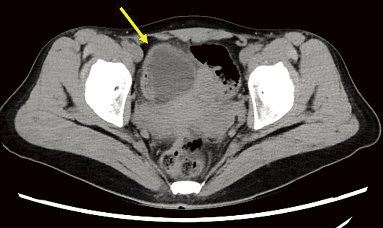
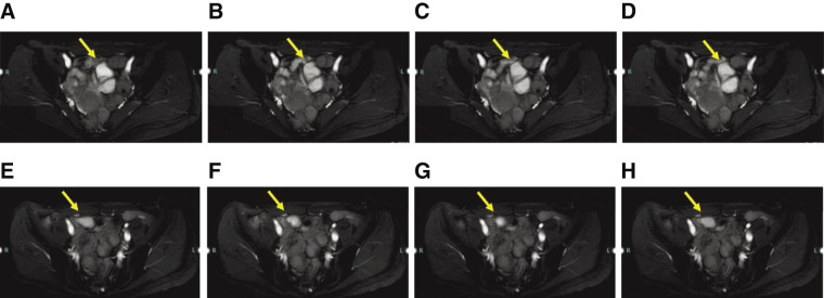
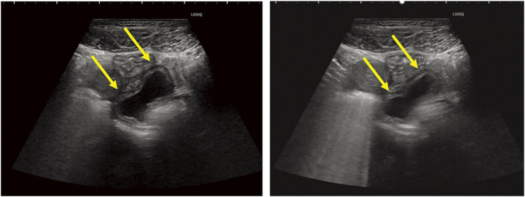
Case presentation: A woman in her thirties visited the emergency department with persistent lower abdominal pain. Physical examination and laboratory tests, including those for tumor markers, were unremarkable. CT revealed a cystic mass near the uterus, and pelvic MRI revealed a cystic lesion that had migrated during follow-up imaging. Cine MRI showed peristaltic movements within the lesion, and abdominal ultrasonography confirmed a cystic structure with wall movements resembling intestinal peristalsis. Based on these findings, the diagnosis of a noncommunicating small bowel duplication cyst was made.The patient underwent a laparoscopic single-port partial resection of the ileum. A cystic lesion located 75 cm proximal to the terminal ileum was excised along with a segment of the small intestine. Histopathological examination revealed a duplicated cyst lined with the small intestinal mucosa, confirming the diagnosis. The postoperative course was uneventful, and the patient was discharged 1 week postoperatively.
Conclusion: This case highlights the utility of cine MRI and ultrasonography in the preoperative diagnosis of small bowel duplication cysts. In particular, cine MRI provides dynamic visualization of peristaltic movements within the cyst, enabling a confident diagnosis. The migration of the cyst observed on serial MRI examinations further corroborated the origin of this duplication. These findings emphasize the importance of advanced imaging modalities in the diagnosis of rare intestinal anomalies. Preoperative diagnosis of small bowel duplication cysts can be significantly enhanced by using cine MRI and ultrasonography to detect peristaltic movements. These modalities offer critical insights that aid timely surgical intervention and improve outcomes.

 求助内容:
求助内容: 应助结果提醒方式:


