{"title":"SIRT1激活通过SIRT1- foxo1 - foxo3自噬途径减少缺氧肾小管上皮细胞纤维化","authors":"Guangyu Wang, Lijuan Zhang, Jiaorong Tan, Fei Li, Yishan Jin, Limei He, Xin Yang","doi":"10.1002/adbi.202400583","DOIUrl":null,"url":null,"abstract":"<p>Heart failure-induced renal tubular epithelial cell fibrosis is an important pathological process that leads to chronic kidney disease. This study is to investigate the regulatory mechanism of over-expression or knock-down SIRT1 gene, alleviating hypoxia-induced HK2 cell fibrosis in heart failure. The focus is on the SIRT1-FoxO1-FoxO3-Autophagy pathway. In vitro experiments are performed by HK2cell line to simulate the normal oxygen state (Normoxia) and the hypoxia state (Hypoxia) caused by heart failure, SIRT1 gene over-expression by transfected vectors, knock-down and Rapamycin (RAPA)-induced cellular autophagy, and the cell models are divided into four subgroups, named control group, oeSIRT1, siSIRT1 and siSIRT1+RAPA. Western blotting (WB), real-time qPCR, immunofluorescence (IF), ELISA, and transmission electron microscopy are used to quantitatively or semi-quantitatively analyze the expression of FoxO1, FoxO3, SIRT1, Beclin1, LC-3, α-SMA, E- Cadherin, and collagen-I in cells or supernatants. It is demonstrated that activation of SIRT1 regulates the expression and activity of FoxO1 and FoxO3, thereby affecting autophagy. This modulation leads to a reduction in HK2 fibrosis markers (α-SMA and E-cadherin) and extracellular matrix deposition (collagen I), which ultimately attenuates renal tubular epithelial cell fibrosis. These findings provide new insights into potential therapeutic strategies for treating heart failure-induced renal tubular epithelial cell fibrosis by targeting the SIRT1-FoxO1-FoxO3-Autophagy pathway.</p>","PeriodicalId":7234,"journal":{"name":"Advanced biology","volume":"9 7","pages":""},"PeriodicalIF":2.6000,"publicationDate":"2025-04-08","publicationTypes":"Journal Article","fieldsOfStudy":null,"isOpenAccess":false,"openAccessPdf":"https://onlinelibrary.wiley.com/doi/epdf/10.1002/adbi.202400583","citationCount":"0","resultStr":"{\"title\":\"Activation of SIRT1 Reduces Renal Tubular Epithelial Cells Fibrosis in Hypoxia Through SIRT1-FoxO1-FoxO3-Autophagy Pathway\",\"authors\":\"Guangyu Wang, Lijuan Zhang, Jiaorong Tan, Fei Li, Yishan Jin, Limei He, Xin Yang\",\"doi\":\"10.1002/adbi.202400583\",\"DOIUrl\":null,\"url\":null,\"abstract\":\"<p>Heart failure-induced renal tubular epithelial cell fibrosis is an important pathological process that leads to chronic kidney disease. This study is to investigate the regulatory mechanism of over-expression or knock-down SIRT1 gene, alleviating hypoxia-induced HK2 cell fibrosis in heart failure. The focus is on the SIRT1-FoxO1-FoxO3-Autophagy pathway. In vitro experiments are performed by HK2cell line to simulate the normal oxygen state (Normoxia) and the hypoxia state (Hypoxia) caused by heart failure, SIRT1 gene over-expression by transfected vectors, knock-down and Rapamycin (RAPA)-induced cellular autophagy, and the cell models are divided into four subgroups, named control group, oeSIRT1, siSIRT1 and siSIRT1+RAPA. Western blotting (WB), real-time qPCR, immunofluorescence (IF), ELISA, and transmission electron microscopy are used to quantitatively or semi-quantitatively analyze the expression of FoxO1, FoxO3, SIRT1, Beclin1, LC-3, α-SMA, E- Cadherin, and collagen-I in cells or supernatants. It is demonstrated that activation of SIRT1 regulates the expression and activity of FoxO1 and FoxO3, thereby affecting autophagy. This modulation leads to a reduction in HK2 fibrosis markers (α-SMA and E-cadherin) and extracellular matrix deposition (collagen I), which ultimately attenuates renal tubular epithelial cell fibrosis. These findings provide new insights into potential therapeutic strategies for treating heart failure-induced renal tubular epithelial cell fibrosis by targeting the SIRT1-FoxO1-FoxO3-Autophagy pathway.</p>\",\"PeriodicalId\":7234,\"journal\":{\"name\":\"Advanced biology\",\"volume\":\"9 7\",\"pages\":\"\"},\"PeriodicalIF\":2.6000,\"publicationDate\":\"2025-04-08\",\"publicationTypes\":\"Journal Article\",\"fieldsOfStudy\":null,\"isOpenAccess\":false,\"openAccessPdf\":\"https://onlinelibrary.wiley.com/doi/epdf/10.1002/adbi.202400583\",\"citationCount\":\"0\",\"resultStr\":null,\"platform\":\"Semanticscholar\",\"paperid\":null,\"PeriodicalName\":\"Advanced biology\",\"FirstCategoryId\":\"99\",\"ListUrlMain\":\"https://advanced.onlinelibrary.wiley.com/doi/10.1002/adbi.202400583\",\"RegionNum\":3,\"RegionCategory\":\"生物学\",\"ArticlePicture\":[],\"TitleCN\":null,\"AbstractTextCN\":null,\"PMCID\":null,\"EPubDate\":\"\",\"PubModel\":\"\",\"JCR\":\"Q3\",\"JCRName\":\"MATERIALS SCIENCE, BIOMATERIALS\",\"Score\":null,\"Total\":0}","platform":"Semanticscholar","paperid":null,"PeriodicalName":"Advanced biology","FirstCategoryId":"99","ListUrlMain":"https://advanced.onlinelibrary.wiley.com/doi/10.1002/adbi.202400583","RegionNum":3,"RegionCategory":"生物学","ArticlePicture":[],"TitleCN":null,"AbstractTextCN":null,"PMCID":null,"EPubDate":"","PubModel":"","JCR":"Q3","JCRName":"MATERIALS SCIENCE, BIOMATERIALS","Score":null,"Total":0}
Activation of SIRT1 Reduces Renal Tubular Epithelial Cells Fibrosis in Hypoxia Through SIRT1-FoxO1-FoxO3-Autophagy Pathway
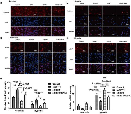
Heart failure-induced renal tubular epithelial cell fibrosis is an important pathological process that leads to chronic kidney disease. This study is to investigate the regulatory mechanism of over-expression or knock-down SIRT1 gene, alleviating hypoxia-induced HK2 cell fibrosis in heart failure. The focus is on the SIRT1-FoxO1-FoxO3-Autophagy pathway. In vitro experiments are performed by HK2cell line to simulate the normal oxygen state (Normoxia) and the hypoxia state (Hypoxia) caused by heart failure, SIRT1 gene over-expression by transfected vectors, knock-down and Rapamycin (RAPA)-induced cellular autophagy, and the cell models are divided into four subgroups, named control group, oeSIRT1, siSIRT1 and siSIRT1+RAPA. Western blotting (WB), real-time qPCR, immunofluorescence (IF), ELISA, and transmission electron microscopy are used to quantitatively or semi-quantitatively analyze the expression of FoxO1, FoxO3, SIRT1, Beclin1, LC-3, α-SMA, E- Cadherin, and collagen-I in cells or supernatants. It is demonstrated that activation of SIRT1 regulates the expression and activity of FoxO1 and FoxO3, thereby affecting autophagy. This modulation leads to a reduction in HK2 fibrosis markers (α-SMA and E-cadherin) and extracellular matrix deposition (collagen I), which ultimately attenuates renal tubular epithelial cell fibrosis. These findings provide new insights into potential therapeutic strategies for treating heart failure-induced renal tubular epithelial cell fibrosis by targeting the SIRT1-FoxO1-FoxO3-Autophagy pathway.


 求助内容:
求助内容: 应助结果提醒方式:


