Elena Tsolaki , Luke Hunter , Adrian H. Chester , Sergio Bertazzo
{"title":"密度依赖性彩色扫描电子显微镜(DDC-SEM)。在钙化组织和视觉冲击研究中的应用","authors":"Elena Tsolaki , Luke Hunter , Adrian H. Chester , Sergio Bertazzo","doi":"10.1016/j.micron.2025.103821","DOIUrl":null,"url":null,"abstract":"<div><div>Scanning Electron Microscopy (SEM) is widely used as a technique for materials characterization. It has also been successfully applied to the imaging of biological samples, providing invaluable insights into the topography, morphology and composition of biological structures. A particular method combining different SEM detectors, named Density-Dependent Coloured SEM (DDC-SEM), has proven to be most useful for the identification and visualization of minerals in soft tissues. The method consists of a manipulation of original greyscale SEM images to produce coloured images that provide both topography and density information for samples with components of different densities. Here we provide a discussion on how to use DDC-SEM to aid the visualization and intuitive understanding of pathological calcification. This method has become popular not only for its scientific improvement of conventional SEM greyscale images, but also for its aesthetical merits.</div></div>","PeriodicalId":18501,"journal":{"name":"Micron","volume":"195 ","pages":"Article 103821"},"PeriodicalIF":2.2000,"publicationDate":"2025-04-01","publicationTypes":"Journal Article","fieldsOfStudy":null,"isOpenAccess":false,"openAccessPdf":"","citationCount":"0","resultStr":"{\"title\":\"Density-dependent colour scanning electron microscopy (DDC-SEM). Applications in the study of calcified tissues and visual impact\",\"authors\":\"Elena Tsolaki , Luke Hunter , Adrian H. Chester , Sergio Bertazzo\",\"doi\":\"10.1016/j.micron.2025.103821\",\"DOIUrl\":null,\"url\":null,\"abstract\":\"<div><div>Scanning Electron Microscopy (SEM) is widely used as a technique for materials characterization. It has also been successfully applied to the imaging of biological samples, providing invaluable insights into the topography, morphology and composition of biological structures. A particular method combining different SEM detectors, named Density-Dependent Coloured SEM (DDC-SEM), has proven to be most useful for the identification and visualization of minerals in soft tissues. The method consists of a manipulation of original greyscale SEM images to produce coloured images that provide both topography and density information for samples with components of different densities. Here we provide a discussion on how to use DDC-SEM to aid the visualization and intuitive understanding of pathological calcification. This method has become popular not only for its scientific improvement of conventional SEM greyscale images, but also for its aesthetical merits.</div></div>\",\"PeriodicalId\":18501,\"journal\":{\"name\":\"Micron\",\"volume\":\"195 \",\"pages\":\"Article 103821\"},\"PeriodicalIF\":2.2000,\"publicationDate\":\"2025-04-01\",\"publicationTypes\":\"Journal Article\",\"fieldsOfStudy\":null,\"isOpenAccess\":false,\"openAccessPdf\":\"\",\"citationCount\":\"0\",\"resultStr\":null,\"platform\":\"Semanticscholar\",\"paperid\":null,\"PeriodicalName\":\"Micron\",\"FirstCategoryId\":\"5\",\"ListUrlMain\":\"https://www.sciencedirect.com/science/article/pii/S0968432825000393\",\"RegionNum\":3,\"RegionCategory\":\"工程技术\",\"ArticlePicture\":[],\"TitleCN\":null,\"AbstractTextCN\":null,\"PMCID\":null,\"EPubDate\":\"\",\"PubModel\":\"\",\"JCR\":\"Q1\",\"JCRName\":\"MICROSCOPY\",\"Score\":null,\"Total\":0}","platform":"Semanticscholar","paperid":null,"PeriodicalName":"Micron","FirstCategoryId":"5","ListUrlMain":"https://www.sciencedirect.com/science/article/pii/S0968432825000393","RegionNum":3,"RegionCategory":"工程技术","ArticlePicture":[],"TitleCN":null,"AbstractTextCN":null,"PMCID":null,"EPubDate":"","PubModel":"","JCR":"Q1","JCRName":"MICROSCOPY","Score":null,"Total":0}
Density-dependent colour scanning electron microscopy (DDC-SEM). Applications in the study of calcified tissues and visual impact
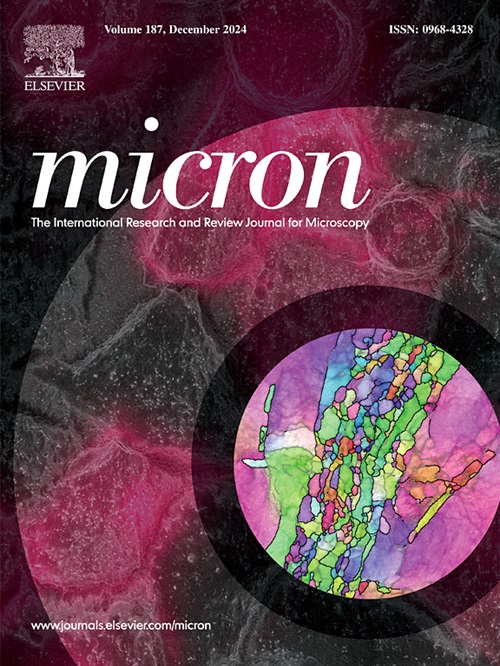
Scanning Electron Microscopy (SEM) is widely used as a technique for materials characterization. It has also been successfully applied to the imaging of biological samples, providing invaluable insights into the topography, morphology and composition of biological structures. A particular method combining different SEM detectors, named Density-Dependent Coloured SEM (DDC-SEM), has proven to be most useful for the identification and visualization of minerals in soft tissues. The method consists of a manipulation of original greyscale SEM images to produce coloured images that provide both topography and density information for samples with components of different densities. Here we provide a discussion on how to use DDC-SEM to aid the visualization and intuitive understanding of pathological calcification. This method has become popular not only for its scientific improvement of conventional SEM greyscale images, but also for its aesthetical merits.
期刊介绍:
Micron is an interdisciplinary forum for all work that involves new applications of microscopy or where advanced microscopy plays a central role. The journal will publish on the design, methods, application, practice or theory of microscopy and microanalysis, including reports on optical, electron-beam, X-ray microtomography, and scanning-probe systems. It also aims at the regular publication of review papers, short communications, as well as thematic issues on contemporary developments in microscopy and microanalysis. The journal embraces original research in which microscopy has contributed significantly to knowledge in biology, life science, nanoscience and nanotechnology, materials science and engineering.