A. M. Arunnagiri, M. Sasikala, N. Ramadass, S. Mullai Venthan
{"title":"高通量显微镜外周血涂片分析贫血筛查的研制。","authors":"A. M. Arunnagiri, M. Sasikala, N. Ramadass, S. Mullai Venthan","doi":"10.1002/jbio.70024","DOIUrl":null,"url":null,"abstract":"<p>The conventional method of screening for anemia requires pathologists to manually examine slides via microscope, a tedious process during health emergencies. This study presents an automated high-throughput optical digital microscope system capable of sequentially scanning and analyzing 10 blood smear slides per batch in under 15 min using a Laplacian-based autofocusing algorithm at 40x magnification. The acquired images are segmented via the YOLOv5 algorithm, and morphological features of red blood cells (RBCs) are classified using a multilayer perceptron (MLP) model. The system achieved 90.6% accuracy, 95% precision, 91% sensitivity, and 94% specificity in classifying anemia subtypes (macrocytic, microcytic, normocytic) and healthy samples. The trained model is integrated into an Android application for real-time geographic mapping of anemic clusters, enabling healthcare workers to prioritize interventions efficiently. This high-throughput approach eliminates the need for immersion oil and manual slide handling, demonstrating significant potential for rapid, scalable anemia screening in resource-limited settings.</p>","PeriodicalId":184,"journal":{"name":"Journal of Biophotonics","volume":"18 8","pages":""},"PeriodicalIF":2.0000,"publicationDate":"2025-04-06","publicationTypes":"Journal Article","fieldsOfStudy":null,"isOpenAccess":false,"openAccessPdf":"","citationCount":"0","resultStr":"{\"title\":\"Development of a High-Throughput Microscope for the Analysis of Peripheral Blood Smears for Anemia Screening\",\"authors\":\"A. M. Arunnagiri, M. Sasikala, N. Ramadass, S. Mullai Venthan\",\"doi\":\"10.1002/jbio.70024\",\"DOIUrl\":null,\"url\":null,\"abstract\":\"<p>The conventional method of screening for anemia requires pathologists to manually examine slides via microscope, a tedious process during health emergencies. This study presents an automated high-throughput optical digital microscope system capable of sequentially scanning and analyzing 10 blood smear slides per batch in under 15 min using a Laplacian-based autofocusing algorithm at 40x magnification. The acquired images are segmented via the YOLOv5 algorithm, and morphological features of red blood cells (RBCs) are classified using a multilayer perceptron (MLP) model. The system achieved 90.6% accuracy, 95% precision, 91% sensitivity, and 94% specificity in classifying anemia subtypes (macrocytic, microcytic, normocytic) and healthy samples. The trained model is integrated into an Android application for real-time geographic mapping of anemic clusters, enabling healthcare workers to prioritize interventions efficiently. This high-throughput approach eliminates the need for immersion oil and manual slide handling, demonstrating significant potential for rapid, scalable anemia screening in resource-limited settings.</p>\",\"PeriodicalId\":184,\"journal\":{\"name\":\"Journal of Biophotonics\",\"volume\":\"18 8\",\"pages\":\"\"},\"PeriodicalIF\":2.0000,\"publicationDate\":\"2025-04-06\",\"publicationTypes\":\"Journal Article\",\"fieldsOfStudy\":null,\"isOpenAccess\":false,\"openAccessPdf\":\"\",\"citationCount\":\"0\",\"resultStr\":null,\"platform\":\"Semanticscholar\",\"paperid\":null,\"PeriodicalName\":\"Journal of Biophotonics\",\"FirstCategoryId\":\"101\",\"ListUrlMain\":\"https://onlinelibrary.wiley.com/doi/10.1002/jbio.70024\",\"RegionNum\":3,\"RegionCategory\":\"物理与天体物理\",\"ArticlePicture\":[],\"TitleCN\":null,\"AbstractTextCN\":null,\"PMCID\":null,\"EPubDate\":\"\",\"PubModel\":\"\",\"JCR\":\"Q3\",\"JCRName\":\"BIOCHEMICAL RESEARCH METHODS\",\"Score\":null,\"Total\":0}","platform":"Semanticscholar","paperid":null,"PeriodicalName":"Journal of Biophotonics","FirstCategoryId":"101","ListUrlMain":"https://onlinelibrary.wiley.com/doi/10.1002/jbio.70024","RegionNum":3,"RegionCategory":"物理与天体物理","ArticlePicture":[],"TitleCN":null,"AbstractTextCN":null,"PMCID":null,"EPubDate":"","PubModel":"","JCR":"Q3","JCRName":"BIOCHEMICAL RESEARCH METHODS","Score":null,"Total":0}
Development of a High-Throughput Microscope for the Analysis of Peripheral Blood Smears for Anemia Screening
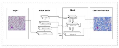
The conventional method of screening for anemia requires pathologists to manually examine slides via microscope, a tedious process during health emergencies. This study presents an automated high-throughput optical digital microscope system capable of sequentially scanning and analyzing 10 blood smear slides per batch in under 15 min using a Laplacian-based autofocusing algorithm at 40x magnification. The acquired images are segmented via the YOLOv5 algorithm, and morphological features of red blood cells (RBCs) are classified using a multilayer perceptron (MLP) model. The system achieved 90.6% accuracy, 95% precision, 91% sensitivity, and 94% specificity in classifying anemia subtypes (macrocytic, microcytic, normocytic) and healthy samples. The trained model is integrated into an Android application for real-time geographic mapping of anemic clusters, enabling healthcare workers to prioritize interventions efficiently. This high-throughput approach eliminates the need for immersion oil and manual slide handling, demonstrating significant potential for rapid, scalable anemia screening in resource-limited settings.
期刊介绍:
The first international journal dedicated to publishing reviews and original articles from this exciting field, the Journal of Biophotonics covers the broad range of research on interactions between light and biological material. The journal offers a platform where the physicist communicates with the biologist and where the clinical practitioner learns about the latest tools for the diagnosis of diseases. As such, the journal is highly interdisciplinary, publishing cutting edge research in the fields of life sciences, medicine, physics, chemistry, and engineering. The coverage extends from fundamental research to specific developments, while also including the latest applications.
 求助内容:
求助内容: 应助结果提醒方式:


