Thamires Mazzola, Geanny Kassia Ferreira Urzêda, Talita Sarah Mazzoni, Marcos José Marques, Hugo Gaêta-Araujo, Marta Miyazawa, Leonardo Amaral Dos Reis, João Adolfo Costa Hanemann
{"title":"影像诊断对模仿牙痛的鼻肌炎的意义。","authors":"Thamires Mazzola, Geanny Kassia Ferreira Urzêda, Talita Sarah Mazzoni, Marcos José Marques, Hugo Gaêta-Araujo, Marta Miyazawa, Leonardo Amaral Dos Reis, João Adolfo Costa Hanemann","doi":"10.5624/isd.20240143","DOIUrl":null,"url":null,"abstract":"<p><p>Nasal myiasis is an infestation by dipterous larvae within the nasal cavity, where they feed on both living tissue and fluid. This condition predominantly occurs in rural areas of tropical countries, where inadequate sanitation and a hot, humid climate create an ideal environment for larvae development. A 57-year-old, otherwise healthy male rural worker presented with a toothache in the region of the maxillary incisors. Imaging studies identified a punctiform radiopaque/hyperdense area near the nasal septum in the left nasal fossa. The larva was surgically excised and sent for histopathological analysis. Histologic sections confirmed the clinical diagnosis, and the patient remained asymptomatic after a 2-month follow-up. Nasal myiasis can mimic the symptoms of a toothache in the anterior region of the maxilla. This condition can affect even immunocompetent patients, and complementary imaging studies may be decisive in diagnosing it.</p>","PeriodicalId":51714,"journal":{"name":"Imaging Science in Dentistry","volume":"55 1","pages":"90-95"},"PeriodicalIF":2.1000,"publicationDate":"2025-03-01","publicationTypes":"Journal Article","fieldsOfStudy":null,"isOpenAccess":false,"openAccessPdf":"https://www.ncbi.nlm.nih.gov/pmc/articles/PMC11966013/pdf/","citationCount":"0","resultStr":"{\"title\":\"The relevance of imaging diagnosis in nasal myiasis mimicking a toothache.\",\"authors\":\"Thamires Mazzola, Geanny Kassia Ferreira Urzêda, Talita Sarah Mazzoni, Marcos José Marques, Hugo Gaêta-Araujo, Marta Miyazawa, Leonardo Amaral Dos Reis, João Adolfo Costa Hanemann\",\"doi\":\"10.5624/isd.20240143\",\"DOIUrl\":null,\"url\":null,\"abstract\":\"<p><p>Nasal myiasis is an infestation by dipterous larvae within the nasal cavity, where they feed on both living tissue and fluid. This condition predominantly occurs in rural areas of tropical countries, where inadequate sanitation and a hot, humid climate create an ideal environment for larvae development. A 57-year-old, otherwise healthy male rural worker presented with a toothache in the region of the maxillary incisors. Imaging studies identified a punctiform radiopaque/hyperdense area near the nasal septum in the left nasal fossa. The larva was surgically excised and sent for histopathological analysis. Histologic sections confirmed the clinical diagnosis, and the patient remained asymptomatic after a 2-month follow-up. Nasal myiasis can mimic the symptoms of a toothache in the anterior region of the maxilla. This condition can affect even immunocompetent patients, and complementary imaging studies may be decisive in diagnosing it.</p>\",\"PeriodicalId\":51714,\"journal\":{\"name\":\"Imaging Science in Dentistry\",\"volume\":\"55 1\",\"pages\":\"90-95\"},\"PeriodicalIF\":2.1000,\"publicationDate\":\"2025-03-01\",\"publicationTypes\":\"Journal Article\",\"fieldsOfStudy\":null,\"isOpenAccess\":false,\"openAccessPdf\":\"https://www.ncbi.nlm.nih.gov/pmc/articles/PMC11966013/pdf/\",\"citationCount\":\"0\",\"resultStr\":null,\"platform\":\"Semanticscholar\",\"paperid\":null,\"PeriodicalName\":\"Imaging Science in Dentistry\",\"FirstCategoryId\":\"1085\",\"ListUrlMain\":\"https://doi.org/10.5624/isd.20240143\",\"RegionNum\":0,\"RegionCategory\":null,\"ArticlePicture\":[],\"TitleCN\":null,\"AbstractTextCN\":null,\"PMCID\":null,\"EPubDate\":\"2024/12/6 0:00:00\",\"PubModel\":\"Epub\",\"JCR\":\"Q3\",\"JCRName\":\"DENTISTRY, ORAL SURGERY & MEDICINE\",\"Score\":null,\"Total\":0}","platform":"Semanticscholar","paperid":null,"PeriodicalName":"Imaging Science in Dentistry","FirstCategoryId":"1085","ListUrlMain":"https://doi.org/10.5624/isd.20240143","RegionNum":0,"RegionCategory":null,"ArticlePicture":[],"TitleCN":null,"AbstractTextCN":null,"PMCID":null,"EPubDate":"2024/12/6 0:00:00","PubModel":"Epub","JCR":"Q3","JCRName":"DENTISTRY, ORAL SURGERY & MEDICINE","Score":null,"Total":0}
The relevance of imaging diagnosis in nasal myiasis mimicking a toothache.
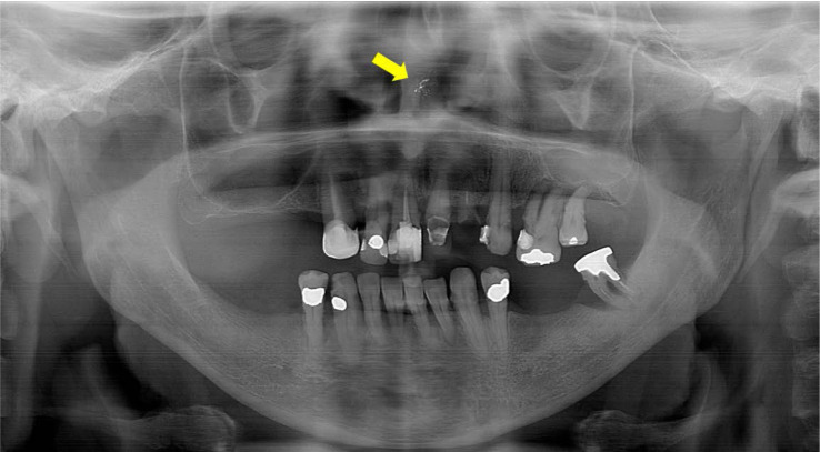
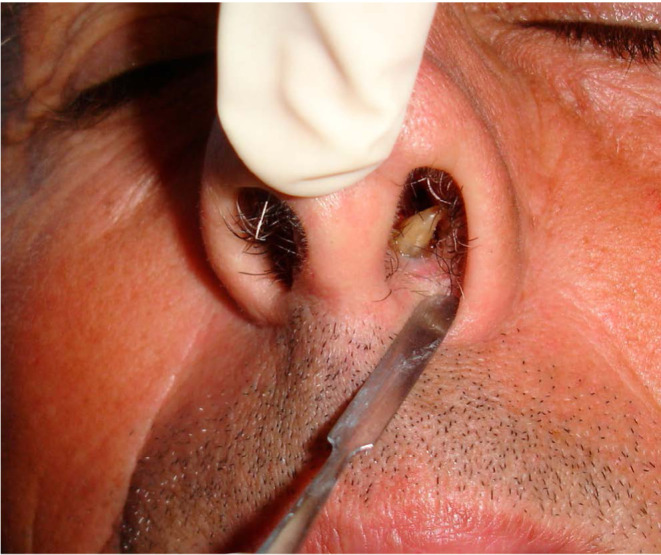
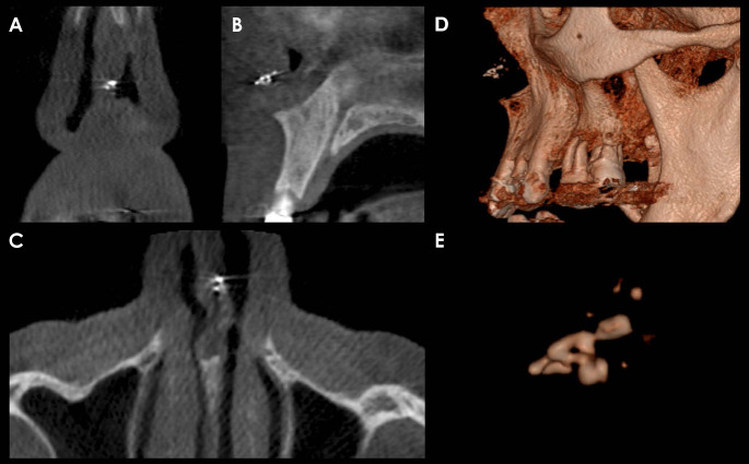
Nasal myiasis is an infestation by dipterous larvae within the nasal cavity, where they feed on both living tissue and fluid. This condition predominantly occurs in rural areas of tropical countries, where inadequate sanitation and a hot, humid climate create an ideal environment for larvae development. A 57-year-old, otherwise healthy male rural worker presented with a toothache in the region of the maxillary incisors. Imaging studies identified a punctiform radiopaque/hyperdense area near the nasal septum in the left nasal fossa. The larva was surgically excised and sent for histopathological analysis. Histologic sections confirmed the clinical diagnosis, and the patient remained asymptomatic after a 2-month follow-up. Nasal myiasis can mimic the symptoms of a toothache in the anterior region of the maxilla. This condition can affect even immunocompetent patients, and complementary imaging studies may be decisive in diagnosing it.

 求助内容:
求助内容: 应助结果提醒方式:


