{"title":"结节性肺淀粉样变的影像学与临床特征。","authors":"Fei Li, Junting Li, Yanyan Li, Danting Shang, Xingyi Hou, Yanli He, Gangfeng Li","doi":"10.1097/RTI.0000000000000830","DOIUrl":null,"url":null,"abstract":"<p><strong>Purpose: </strong>To investigate the clinical and computed tomography (CT) features of nodular pulmonary amyloidosis (NPA) to enhance our understanding of the disease and improve the ability to differentiate it from other similar conditions.</p><p><strong>Materials and methods: </strong>A retrospective analysis was conducted on the clinical data, chest CT imaging findings, and pathologic characteristics of 13 patients with NPA in our hospital from April 2014 to April 2024. All 13 patients underwent chest CT plain scan examination. The basic data, medical history, clinical manifestations, and lung lesion features on chest CT imaging were analyzed and summarized.</p><p><strong>Results: </strong>Among the 13 patients, there were 3 males (23.08%) and 10 females (76.92%). Their ages ranged from 37 to 68 years, with a mean age of (57.85±8.40) years and a median age of 59 years. Three (23.08%) patients had cough and sputum, while the others (76.92%) had no clinical symptoms. Before surgery, 6 patients underwent chest CT scans, and NPA changes in size, shape, and density were observed. Six cases (46.15%) were located in the left lung (4 in the upper lobe and 2 in the lower lobe), and 7 cases (53.85%) in the right lung (3 in the upper lobe, 2 in the middle lobe, and 2 in the lower lobe). Seven cases (53.85%) of NPA were round or oval, while 6 cases (46.15%) were irregularly shaped. Out of the NPA cases, 11 (84.62%) were solid nodules with well-defined boundaries, including 2 cases of solid nodules with surrounding calcification. In addition, 2 cases presented as solid nodules with cavities. Ten cases (76.92%) had multiple cystic lesions in the bilateral lungs, in which 7 cases had more than 10 cysts with obvious cyst walls, and 1 case showed a solid nodule on the cyst wall. During the postoperative follow-up, 1 patient experienced an increase in the size of the original nodule and the appearance of new solid nodules. Subsequent surgery revealed mucosal-associated lymphoid tissue lymphoma (MALT). The remaining patients were followed up regularly, and their conditions remained stable.</p><p><strong>Conclusions: </strong>NPA is more common in middle-aged and elderly people and is more likely to occur in women. Most cases are asymptomatic, and bilateral lungs can be involved. For nodules with multiple pulmonary cysts found by chest CT, the possibility of NPA should be considered, and further histopathologic examination is needed to confirm the diagnosis. Most patients with NPA have a good long-term prognosis after surgical resection, but some patients require further investigation and close follow-up due to underlying causes.</p>","PeriodicalId":49974,"journal":{"name":"Journal of Thoracic Imaging","volume":" ","pages":""},"PeriodicalIF":1.9000,"publicationDate":"2025-09-01","publicationTypes":"Journal Article","fieldsOfStudy":null,"isOpenAccess":false,"openAccessPdf":"https://www.ncbi.nlm.nih.gov/pmc/articles/PMC12369498/pdf/","citationCount":"0","resultStr":"{\"title\":\"Imaging and Clinical Features of Nodular Pulmonary Amyloidosis.\",\"authors\":\"Fei Li, Junting Li, Yanyan Li, Danting Shang, Xingyi Hou, Yanli He, Gangfeng Li\",\"doi\":\"10.1097/RTI.0000000000000830\",\"DOIUrl\":null,\"url\":null,\"abstract\":\"<p><strong>Purpose: </strong>To investigate the clinical and computed tomography (CT) features of nodular pulmonary amyloidosis (NPA) to enhance our understanding of the disease and improve the ability to differentiate it from other similar conditions.</p><p><strong>Materials and methods: </strong>A retrospective analysis was conducted on the clinical data, chest CT imaging findings, and pathologic characteristics of 13 patients with NPA in our hospital from April 2014 to April 2024. All 13 patients underwent chest CT plain scan examination. The basic data, medical history, clinical manifestations, and lung lesion features on chest CT imaging were analyzed and summarized.</p><p><strong>Results: </strong>Among the 13 patients, there were 3 males (23.08%) and 10 females (76.92%). Their ages ranged from 37 to 68 years, with a mean age of (57.85±8.40) years and a median age of 59 years. Three (23.08%) patients had cough and sputum, while the others (76.92%) had no clinical symptoms. Before surgery, 6 patients underwent chest CT scans, and NPA changes in size, shape, and density were observed. Six cases (46.15%) were located in the left lung (4 in the upper lobe and 2 in the lower lobe), and 7 cases (53.85%) in the right lung (3 in the upper lobe, 2 in the middle lobe, and 2 in the lower lobe). Seven cases (53.85%) of NPA were round or oval, while 6 cases (46.15%) were irregularly shaped. Out of the NPA cases, 11 (84.62%) were solid nodules with well-defined boundaries, including 2 cases of solid nodules with surrounding calcification. In addition, 2 cases presented as solid nodules with cavities. Ten cases (76.92%) had multiple cystic lesions in the bilateral lungs, in which 7 cases had more than 10 cysts with obvious cyst walls, and 1 case showed a solid nodule on the cyst wall. During the postoperative follow-up, 1 patient experienced an increase in the size of the original nodule and the appearance of new solid nodules. Subsequent surgery revealed mucosal-associated lymphoid tissue lymphoma (MALT). The remaining patients were followed up regularly, and their conditions remained stable.</p><p><strong>Conclusions: </strong>NPA is more common in middle-aged and elderly people and is more likely to occur in women. Most cases are asymptomatic, and bilateral lungs can be involved. For nodules with multiple pulmonary cysts found by chest CT, the possibility of NPA should be considered, and further histopathologic examination is needed to confirm the diagnosis. Most patients with NPA have a good long-term prognosis after surgical resection, but some patients require further investigation and close follow-up due to underlying causes.</p>\",\"PeriodicalId\":49974,\"journal\":{\"name\":\"Journal of Thoracic Imaging\",\"volume\":\" \",\"pages\":\"\"},\"PeriodicalIF\":1.9000,\"publicationDate\":\"2025-09-01\",\"publicationTypes\":\"Journal Article\",\"fieldsOfStudy\":null,\"isOpenAccess\":false,\"openAccessPdf\":\"https://www.ncbi.nlm.nih.gov/pmc/articles/PMC12369498/pdf/\",\"citationCount\":\"0\",\"resultStr\":null,\"platform\":\"Semanticscholar\",\"paperid\":null,\"PeriodicalName\":\"Journal of Thoracic Imaging\",\"FirstCategoryId\":\"3\",\"ListUrlMain\":\"https://doi.org/10.1097/RTI.0000000000000830\",\"RegionNum\":4,\"RegionCategory\":\"医学\",\"ArticlePicture\":[],\"TitleCN\":null,\"AbstractTextCN\":null,\"PMCID\":null,\"EPubDate\":\"\",\"PubModel\":\"\",\"JCR\":\"Q3\",\"JCRName\":\"RADIOLOGY, NUCLEAR MEDICINE & MEDICAL IMAGING\",\"Score\":null,\"Total\":0}","platform":"Semanticscholar","paperid":null,"PeriodicalName":"Journal of Thoracic Imaging","FirstCategoryId":"3","ListUrlMain":"https://doi.org/10.1097/RTI.0000000000000830","RegionNum":4,"RegionCategory":"医学","ArticlePicture":[],"TitleCN":null,"AbstractTextCN":null,"PMCID":null,"EPubDate":"","PubModel":"","JCR":"Q3","JCRName":"RADIOLOGY, NUCLEAR MEDICINE & MEDICAL IMAGING","Score":null,"Total":0}
Imaging and Clinical Features of Nodular Pulmonary Amyloidosis.
Purpose: To investigate the clinical and computed tomography (CT) features of nodular pulmonary amyloidosis (NPA) to enhance our understanding of the disease and improve the ability to differentiate it from other similar conditions.
Materials and methods: A retrospective analysis was conducted on the clinical data, chest CT imaging findings, and pathologic characteristics of 13 patients with NPA in our hospital from April 2014 to April 2024. All 13 patients underwent chest CT plain scan examination. The basic data, medical history, clinical manifestations, and lung lesion features on chest CT imaging were analyzed and summarized.
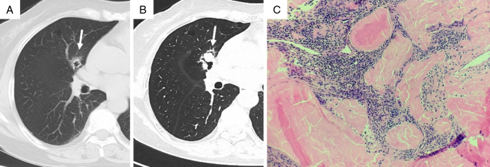
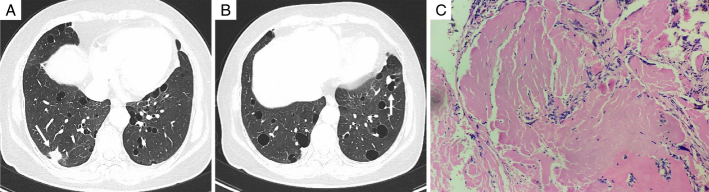
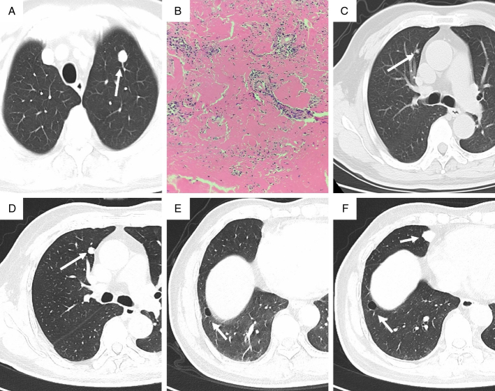
Results: Among the 13 patients, there were 3 males (23.08%) and 10 females (76.92%). Their ages ranged from 37 to 68 years, with a mean age of (57.85±8.40) years and a median age of 59 years. Three (23.08%) patients had cough and sputum, while the others (76.92%) had no clinical symptoms. Before surgery, 6 patients underwent chest CT scans, and NPA changes in size, shape, and density were observed. Six cases (46.15%) were located in the left lung (4 in the upper lobe and 2 in the lower lobe), and 7 cases (53.85%) in the right lung (3 in the upper lobe, 2 in the middle lobe, and 2 in the lower lobe). Seven cases (53.85%) of NPA were round or oval, while 6 cases (46.15%) were irregularly shaped. Out of the NPA cases, 11 (84.62%) were solid nodules with well-defined boundaries, including 2 cases of solid nodules with surrounding calcification. In addition, 2 cases presented as solid nodules with cavities. Ten cases (76.92%) had multiple cystic lesions in the bilateral lungs, in which 7 cases had more than 10 cysts with obvious cyst walls, and 1 case showed a solid nodule on the cyst wall. During the postoperative follow-up, 1 patient experienced an increase in the size of the original nodule and the appearance of new solid nodules. Subsequent surgery revealed mucosal-associated lymphoid tissue lymphoma (MALT). The remaining patients were followed up regularly, and their conditions remained stable.
Conclusions: NPA is more common in middle-aged and elderly people and is more likely to occur in women. Most cases are asymptomatic, and bilateral lungs can be involved. For nodules with multiple pulmonary cysts found by chest CT, the possibility of NPA should be considered, and further histopathologic examination is needed to confirm the diagnosis. Most patients with NPA have a good long-term prognosis after surgical resection, but some patients require further investigation and close follow-up due to underlying causes.
期刊介绍:
Journal of Thoracic Imaging (JTI) provides authoritative information on all aspects of the use of imaging techniques in the diagnosis of cardiac and pulmonary diseases. Original articles and analytical reviews published in this timely journal provide the very latest thinking of leading experts concerning the use of chest radiography, computed tomography, magnetic resonance imaging, positron emission tomography, ultrasound, and all other promising imaging techniques in cardiopulmonary radiology.
Official Journal of the Society of Thoracic Radiology:
Japanese Society of Thoracic Radiology
Korean Society of Thoracic Radiology
European Society of Thoracic Imaging.

 求助内容:
求助内容: 应助结果提醒方式:


