{"title":"毫安电流对牙种植体周围骨移植体锥形束ct评价的影响。","authors":"Henrique Mateus Alves Felizardo, Bruna Silveira Troca, Polyane Mazucatto Queiroz, Hugo Gaêta-Araujo","doi":"10.5624/isd.20240214","DOIUrl":null,"url":null,"abstract":"<p><strong>Purpose: </strong>Bone grafts can be challenging to assess on cone-beam computed tomography (CBCT) examinations due to their discreet appearance and the potential introduction of metallic artifacts from implant screws. This study aimed to evaluate the effect of CBCT milliamperage (mA) on detecting bone graft dehiscence adjacent to titanium (Ti) and zirconia (Zr) implants.</p><p><strong>Materials and methods: </strong>Twenty Ti and 20 Zr implants were installed in bovine rib blocks. Gaps of at least 2 mm were created between the implant and the bone and filled with particulate autogenous bone grafts. In half of the blocks, the gap was completely filled, while in the other half, the grafting material was removed up to the third implant thread. CBCT images were acquired at 4, 6.3, and 10 mA and evaluated by 5 observers to detect bone graft dehiscence. The area under the receiver operating characteristic curve, accuracy, sensitivity, and specificity were calculated. These values were then compared across various dental implant materials and mA levels using 2-way analysis of variance with a significance level of 5%.</p><p><strong>Results: </strong>No statistically significant differences were observed in the diagnostic values for bone graft dehiscence between implant types (<i>P</i>>0.05) or mA settings (<i>P</i>>0.05).</p><p><strong>Conclusion: </strong>Although a protocol with lower radiation exposure (that is, lower mA) could be employed, the use of CBCT for evaluating bone graft dehiscence adjacent to different types of dental implants should be approached with caution.</p>","PeriodicalId":51714,"journal":{"name":"Imaging Science in Dentistry","volume":"55 1","pages":"48-55"},"PeriodicalIF":2.1000,"publicationDate":"2025-03-01","publicationTypes":"Journal Article","fieldsOfStudy":null,"isOpenAccess":false,"openAccessPdf":"https://www.ncbi.nlm.nih.gov/pmc/articles/PMC11966024/pdf/","citationCount":"0","resultStr":"{\"title\":\"Effect of milliamperage on cone-beam computed tomography evaluation of bone grafts around dental implants.\",\"authors\":\"Henrique Mateus Alves Felizardo, Bruna Silveira Troca, Polyane Mazucatto Queiroz, Hugo Gaêta-Araujo\",\"doi\":\"10.5624/isd.20240214\",\"DOIUrl\":null,\"url\":null,\"abstract\":\"<p><strong>Purpose: </strong>Bone grafts can be challenging to assess on cone-beam computed tomography (CBCT) examinations due to their discreet appearance and the potential introduction of metallic artifacts from implant screws. This study aimed to evaluate the effect of CBCT milliamperage (mA) on detecting bone graft dehiscence adjacent to titanium (Ti) and zirconia (Zr) implants.</p><p><strong>Materials and methods: </strong>Twenty Ti and 20 Zr implants were installed in bovine rib blocks. Gaps of at least 2 mm were created between the implant and the bone and filled with particulate autogenous bone grafts. In half of the blocks, the gap was completely filled, while in the other half, the grafting material was removed up to the third implant thread. CBCT images were acquired at 4, 6.3, and 10 mA and evaluated by 5 observers to detect bone graft dehiscence. The area under the receiver operating characteristic curve, accuracy, sensitivity, and specificity were calculated. These values were then compared across various dental implant materials and mA levels using 2-way analysis of variance with a significance level of 5%.</p><p><strong>Results: </strong>No statistically significant differences were observed in the diagnostic values for bone graft dehiscence between implant types (<i>P</i>>0.05) or mA settings (<i>P</i>>0.05).</p><p><strong>Conclusion: </strong>Although a protocol with lower radiation exposure (that is, lower mA) could be employed, the use of CBCT for evaluating bone graft dehiscence adjacent to different types of dental implants should be approached with caution.</p>\",\"PeriodicalId\":51714,\"journal\":{\"name\":\"Imaging Science in Dentistry\",\"volume\":\"55 1\",\"pages\":\"48-55\"},\"PeriodicalIF\":2.1000,\"publicationDate\":\"2025-03-01\",\"publicationTypes\":\"Journal Article\",\"fieldsOfStudy\":null,\"isOpenAccess\":false,\"openAccessPdf\":\"https://www.ncbi.nlm.nih.gov/pmc/articles/PMC11966024/pdf/\",\"citationCount\":\"0\",\"resultStr\":null,\"platform\":\"Semanticscholar\",\"paperid\":null,\"PeriodicalName\":\"Imaging Science in Dentistry\",\"FirstCategoryId\":\"1085\",\"ListUrlMain\":\"https://doi.org/10.5624/isd.20240214\",\"RegionNum\":0,\"RegionCategory\":null,\"ArticlePicture\":[],\"TitleCN\":null,\"AbstractTextCN\":null,\"PMCID\":null,\"EPubDate\":\"2025/3/10 0:00:00\",\"PubModel\":\"Epub\",\"JCR\":\"Q3\",\"JCRName\":\"DENTISTRY, ORAL SURGERY & MEDICINE\",\"Score\":null,\"Total\":0}","platform":"Semanticscholar","paperid":null,"PeriodicalName":"Imaging Science in Dentistry","FirstCategoryId":"1085","ListUrlMain":"https://doi.org/10.5624/isd.20240214","RegionNum":0,"RegionCategory":null,"ArticlePicture":[],"TitleCN":null,"AbstractTextCN":null,"PMCID":null,"EPubDate":"2025/3/10 0:00:00","PubModel":"Epub","JCR":"Q3","JCRName":"DENTISTRY, ORAL SURGERY & MEDICINE","Score":null,"Total":0}
Effect of milliamperage on cone-beam computed tomography evaluation of bone grafts around dental implants.
Purpose: Bone grafts can be challenging to assess on cone-beam computed tomography (CBCT) examinations due to their discreet appearance and the potential introduction of metallic artifacts from implant screws. This study aimed to evaluate the effect of CBCT milliamperage (mA) on detecting bone graft dehiscence adjacent to titanium (Ti) and zirconia (Zr) implants.
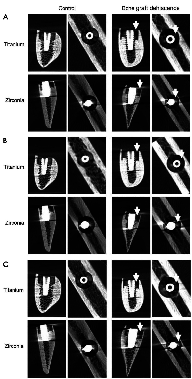
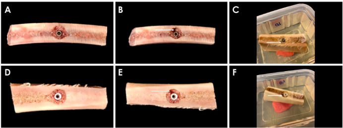
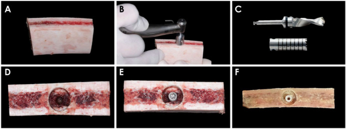
Materials and methods: Twenty Ti and 20 Zr implants were installed in bovine rib blocks. Gaps of at least 2 mm were created between the implant and the bone and filled with particulate autogenous bone grafts. In half of the blocks, the gap was completely filled, while in the other half, the grafting material was removed up to the third implant thread. CBCT images were acquired at 4, 6.3, and 10 mA and evaluated by 5 observers to detect bone graft dehiscence. The area under the receiver operating characteristic curve, accuracy, sensitivity, and specificity were calculated. These values were then compared across various dental implant materials and mA levels using 2-way analysis of variance with a significance level of 5%.
Results: No statistically significant differences were observed in the diagnostic values for bone graft dehiscence between implant types (P>0.05) or mA settings (P>0.05).
Conclusion: Although a protocol with lower radiation exposure (that is, lower mA) could be employed, the use of CBCT for evaluating bone graft dehiscence adjacent to different types of dental implants should be approached with caution.

 求助内容:
求助内容: 应助结果提醒方式:


