Abdelwahab Aleshawi, Mohammad Ali Al Qudah, Hamad Alazmi, Ahmed Al Sharie, Haya Sufyan Elnagar Jnr, Hosni Alzoubi, Saad Almutairi, Seren Al Beiruti, Rami Al-Dwairi
{"title":"假性角膜病眼球内人工晶体上出现大面积视觉显著的假性角膜外沉积物的异常表现:启示与意义。","authors":"Abdelwahab Aleshawi, Mohammad Ali Al Qudah, Hamad Alazmi, Ahmed Al Sharie, Haya Sufyan Elnagar Jnr, Hosni Alzoubi, Saad Almutairi, Seren Al Beiruti, Rami Al-Dwairi","doi":"10.2147/OPTO.S517593","DOIUrl":null,"url":null,"abstract":"<p><strong>Objective: </strong>The aim of this report is to present a rare case of visually significant pseudoexfoliative deposition on an intraocular lens implant in a pseudophakic eye and describe the clinical characteristics, surgical approach, and histopathological characteristics.</p><p><strong>Case presentation: </strong>We present the case of a 61-year-old male with a history of cataract surgery in the left eye who presented with blurry vision bilaterally. Examination of the left eye revealed a centered posterior-chamber intraocular lens implant (IOL) with extensive, visually significant pseudoexfoliative plaques growing over the entire IOL. The decision was made to perform right eye phacoemulsification and IOL implantation and left eye IOL exchange. The tissue specimens were subsequently processed using the classical histological technique of paraffin embedding. Intraocular surgery improved the visual acuity from 6/30 in the left and 6/60 in the right eye to 6/6 in both eyes at 3 months postoperative. The extracted implant was examined under a microscope and showed amyloid-positive deposits.</p><p><strong>Conclusion: </strong>This case highlights the importance of comprehensive examination for pseudoexfoliation, even in patients with pseudophakia. Visually significant PEX deposition was first reported and should be managed with caution.</p>","PeriodicalId":43701,"journal":{"name":"Clinical Optometry","volume":"17 ","pages":"127-131"},"PeriodicalIF":1.8000,"publicationDate":"2025-03-31","publicationTypes":"Journal Article","fieldsOfStudy":null,"isOpenAccess":false,"openAccessPdf":"https://www.ncbi.nlm.nih.gov/pmc/articles/PMC11970284/pdf/","citationCount":"0","resultStr":"{\"title\":\"Unusual Presentation of Extensive Visually Significant Pseudoexfoliative Deposits on an Intraocular Lens Implant in a Pseudophakic Eye: Insights and Implications.\",\"authors\":\"Abdelwahab Aleshawi, Mohammad Ali Al Qudah, Hamad Alazmi, Ahmed Al Sharie, Haya Sufyan Elnagar Jnr, Hosni Alzoubi, Saad Almutairi, Seren Al Beiruti, Rami Al-Dwairi\",\"doi\":\"10.2147/OPTO.S517593\",\"DOIUrl\":null,\"url\":null,\"abstract\":\"<p><strong>Objective: </strong>The aim of this report is to present a rare case of visually significant pseudoexfoliative deposition on an intraocular lens implant in a pseudophakic eye and describe the clinical characteristics, surgical approach, and histopathological characteristics.</p><p><strong>Case presentation: </strong>We present the case of a 61-year-old male with a history of cataract surgery in the left eye who presented with blurry vision bilaterally. Examination of the left eye revealed a centered posterior-chamber intraocular lens implant (IOL) with extensive, visually significant pseudoexfoliative plaques growing over the entire IOL. The decision was made to perform right eye phacoemulsification and IOL implantation and left eye IOL exchange. The tissue specimens were subsequently processed using the classical histological technique of paraffin embedding. Intraocular surgery improved the visual acuity from 6/30 in the left and 6/60 in the right eye to 6/6 in both eyes at 3 months postoperative. The extracted implant was examined under a microscope and showed amyloid-positive deposits.</p><p><strong>Conclusion: </strong>This case highlights the importance of comprehensive examination for pseudoexfoliation, even in patients with pseudophakia. Visually significant PEX deposition was first reported and should be managed with caution.</p>\",\"PeriodicalId\":43701,\"journal\":{\"name\":\"Clinical Optometry\",\"volume\":\"17 \",\"pages\":\"127-131\"},\"PeriodicalIF\":1.8000,\"publicationDate\":\"2025-03-31\",\"publicationTypes\":\"Journal Article\",\"fieldsOfStudy\":null,\"isOpenAccess\":false,\"openAccessPdf\":\"https://www.ncbi.nlm.nih.gov/pmc/articles/PMC11970284/pdf/\",\"citationCount\":\"0\",\"resultStr\":null,\"platform\":\"Semanticscholar\",\"paperid\":null,\"PeriodicalName\":\"Clinical Optometry\",\"FirstCategoryId\":\"1085\",\"ListUrlMain\":\"https://doi.org/10.2147/OPTO.S517593\",\"RegionNum\":0,\"RegionCategory\":null,\"ArticlePicture\":[],\"TitleCN\":null,\"AbstractTextCN\":null,\"PMCID\":null,\"EPubDate\":\"2025/1/1 0:00:00\",\"PubModel\":\"eCollection\",\"JCR\":\"Q3\",\"JCRName\":\"OPHTHALMOLOGY\",\"Score\":null,\"Total\":0}","platform":"Semanticscholar","paperid":null,"PeriodicalName":"Clinical Optometry","FirstCategoryId":"1085","ListUrlMain":"https://doi.org/10.2147/OPTO.S517593","RegionNum":0,"RegionCategory":null,"ArticlePicture":[],"TitleCN":null,"AbstractTextCN":null,"PMCID":null,"EPubDate":"2025/1/1 0:00:00","PubModel":"eCollection","JCR":"Q3","JCRName":"OPHTHALMOLOGY","Score":null,"Total":0}
Unusual Presentation of Extensive Visually Significant Pseudoexfoliative Deposits on an Intraocular Lens Implant in a Pseudophakic Eye: Insights and Implications.
Objective: The aim of this report is to present a rare case of visually significant pseudoexfoliative deposition on an intraocular lens implant in a pseudophakic eye and describe the clinical characteristics, surgical approach, and histopathological characteristics.
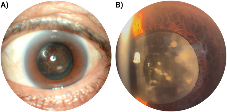
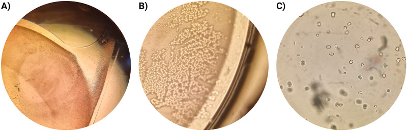
Case presentation: We present the case of a 61-year-old male with a history of cataract surgery in the left eye who presented with blurry vision bilaterally. Examination of the left eye revealed a centered posterior-chamber intraocular lens implant (IOL) with extensive, visually significant pseudoexfoliative plaques growing over the entire IOL. The decision was made to perform right eye phacoemulsification and IOL implantation and left eye IOL exchange. The tissue specimens were subsequently processed using the classical histological technique of paraffin embedding. Intraocular surgery improved the visual acuity from 6/30 in the left and 6/60 in the right eye to 6/6 in both eyes at 3 months postoperative. The extracted implant was examined under a microscope and showed amyloid-positive deposits.
Conclusion: This case highlights the importance of comprehensive examination for pseudoexfoliation, even in patients with pseudophakia. Visually significant PEX deposition was first reported and should be managed with caution.
期刊介绍:
Clinical Optometry is an international, peer-reviewed, open access journal focusing on clinical optometry. All aspects of patient care are addressed within the journal as well as the practice of optometry including economic and business analyses. Basic and clinical research papers are published that cover all aspects of optics, refraction and its application to the theory and practice of optometry. Specific topics covered in the journal include: Theoretical and applied optics, Delivery of patient care in optometry practice, Refraction and correction of errors, Screening and preventative aspects of eye disease, Extended clinical roles for optometrists including shared care and provision of medications, Teaching and training optometrists, International aspects of optometry, Business practice, Patient adherence, quality of life, satisfaction, Health economic evaluations.

 求助内容:
求助内容: 应助结果提醒方式:


