{"title":"幼年特发性关节炎关节的角度评估。","authors":"Sudip Banerjee, Atanu Adak, Debadyuti Dutta, Partha Pratim Pan, Manab Nandy, Avijit Hazra, Rakesh K Mondal","doi":"10.1515/rir-2025-0001","DOIUrl":null,"url":null,"abstract":"<p><strong>Background: </strong>Joint deformities in juvenile idiopathic arthritis (JIA) are most common in children, are not defined in term of angular measurements. The study was aimed to evaluate the joint deformities in angular deviation of the afected joints in JIA patients.</p><p><strong>Methods: </strong>This cross-sectional study was conducted at Pediatric Rheumatology Clinic, North Bengal Medical College, West Bengal. The children aged 2-16 years diagnosed with JIA according to the International League of Associations for Rheumatology (ILAR) criteria were included in the study. Patients with co-morbid disease, hemodynamic instability, and other acute conditions were excluded. Angular measurements were performed using goniometer.</p><p><strong>Results: </strong>The mean age of children was (8.05 ± 3.20) years of which 57.5% was male and the disease duration associated with the deformities in JIA. The prevalent subtypes of JIA were Oligoarticular JIA (oligoJIA)(40%), followed by polyarticular JIA (pJIA) (35%) and systemic-onset JIA (sJIA) (12.5%). The commonly involved joint were knee (40%), followed by small joint of hand (32.5%), ankle (30%), wrist and foot (17.5% each), elbow (12.5%) and cervical joint (7.5%). In pJIA, duration of disease significantly (<i>P</i> = 0.017) associated with the number of affected joints. Mostly, wrist, knee and ankle deformities were observed in oligoJIA, pJIA and sJIA. The angular deviation (mean ± SD) of right and left knee were (2° ± 4.16°) and (1.87° ± 5.12°) in oligoJIA, (13.36° ± 17.03°) and (12.5° ± 15.08°) in pJIA and (3° ± 6.71°) and (2.4° ± 5.37°) in sJIA. Right ankle angular deviation were (2.62° ± 5.06), (5.43° ± 8.21°) and 4° ± 8.94° respectively in oligoJIA, pJIA and sJIA. The angular deviation of right and left wrist were (1.25° ± 3.41°) and (0.94° ± 3.75°) in oligoJIA, (4.07° ± 8.93°) and (4.14° ± 9.36°) in pJIA and (2.45° ± 5.37°) and (2° ± 4.47°) in sJIA.</p><p><strong>Conclusion: </strong>This study is the first study from India to quantify the angular deviation of deformed joints in JIA. Angular deviation could serve as a valuable parameter for monitoring disease progression across various JIA subtypes.</p>","PeriodicalId":74736,"journal":{"name":"Rheumatology and immunology research","volume":"6 1","pages":"1-6"},"PeriodicalIF":2.5000,"publicationDate":"2025-04-02","publicationTypes":"Journal Article","fieldsOfStudy":null,"isOpenAccess":false,"openAccessPdf":"https://www.ncbi.nlm.nih.gov/pmc/articles/PMC11966197/pdf/","citationCount":"0","resultStr":"{\"title\":\"Angular assessment of joints in juvenile idiopathic arthritis.\",\"authors\":\"Sudip Banerjee, Atanu Adak, Debadyuti Dutta, Partha Pratim Pan, Manab Nandy, Avijit Hazra, Rakesh K Mondal\",\"doi\":\"10.1515/rir-2025-0001\",\"DOIUrl\":null,\"url\":null,\"abstract\":\"<p><strong>Background: </strong>Joint deformities in juvenile idiopathic arthritis (JIA) are most common in children, are not defined in term of angular measurements. The study was aimed to evaluate the joint deformities in angular deviation of the afected joints in JIA patients.</p><p><strong>Methods: </strong>This cross-sectional study was conducted at Pediatric Rheumatology Clinic, North Bengal Medical College, West Bengal. The children aged 2-16 years diagnosed with JIA according to the International League of Associations for Rheumatology (ILAR) criteria were included in the study. Patients with co-morbid disease, hemodynamic instability, and other acute conditions were excluded. Angular measurements were performed using goniometer.</p><p><strong>Results: </strong>The mean age of children was (8.05 ± 3.20) years of which 57.5% was male and the disease duration associated with the deformities in JIA. The prevalent subtypes of JIA were Oligoarticular JIA (oligoJIA)(40%), followed by polyarticular JIA (pJIA) (35%) and systemic-onset JIA (sJIA) (12.5%). The commonly involved joint were knee (40%), followed by small joint of hand (32.5%), ankle (30%), wrist and foot (17.5% each), elbow (12.5%) and cervical joint (7.5%). In pJIA, duration of disease significantly (<i>P</i> = 0.017) associated with the number of affected joints. Mostly, wrist, knee and ankle deformities were observed in oligoJIA, pJIA and sJIA. The angular deviation (mean ± SD) of right and left knee were (2° ± 4.16°) and (1.87° ± 5.12°) in oligoJIA, (13.36° ± 17.03°) and (12.5° ± 15.08°) in pJIA and (3° ± 6.71°) and (2.4° ± 5.37°) in sJIA. Right ankle angular deviation were (2.62° ± 5.06), (5.43° ± 8.21°) and 4° ± 8.94° respectively in oligoJIA, pJIA and sJIA. The angular deviation of right and left wrist were (1.25° ± 3.41°) and (0.94° ± 3.75°) in oligoJIA, (4.07° ± 8.93°) and (4.14° ± 9.36°) in pJIA and (2.45° ± 5.37°) and (2° ± 4.47°) in sJIA.</p><p><strong>Conclusion: </strong>This study is the first study from India to quantify the angular deviation of deformed joints in JIA. Angular deviation could serve as a valuable parameter for monitoring disease progression across various JIA subtypes.</p>\",\"PeriodicalId\":74736,\"journal\":{\"name\":\"Rheumatology and immunology research\",\"volume\":\"6 1\",\"pages\":\"1-6\"},\"PeriodicalIF\":2.5000,\"publicationDate\":\"2025-04-02\",\"publicationTypes\":\"Journal Article\",\"fieldsOfStudy\":null,\"isOpenAccess\":false,\"openAccessPdf\":\"https://www.ncbi.nlm.nih.gov/pmc/articles/PMC11966197/pdf/\",\"citationCount\":\"0\",\"resultStr\":null,\"platform\":\"Semanticscholar\",\"paperid\":null,\"PeriodicalName\":\"Rheumatology and immunology research\",\"FirstCategoryId\":\"1085\",\"ListUrlMain\":\"https://doi.org/10.1515/rir-2025-0001\",\"RegionNum\":0,\"RegionCategory\":null,\"ArticlePicture\":[],\"TitleCN\":null,\"AbstractTextCN\":null,\"PMCID\":null,\"EPubDate\":\"2025/3/1 0:00:00\",\"PubModel\":\"eCollection\",\"JCR\":\"\",\"JCRName\":\"\",\"Score\":null,\"Total\":0}","platform":"Semanticscholar","paperid":null,"PeriodicalName":"Rheumatology and immunology research","FirstCategoryId":"1085","ListUrlMain":"https://doi.org/10.1515/rir-2025-0001","RegionNum":0,"RegionCategory":null,"ArticlePicture":[],"TitleCN":null,"AbstractTextCN":null,"PMCID":null,"EPubDate":"2025/3/1 0:00:00","PubModel":"eCollection","JCR":"","JCRName":"","Score":null,"Total":0}
Angular assessment of joints in juvenile idiopathic arthritis.
Background: Joint deformities in juvenile idiopathic arthritis (JIA) are most common in children, are not defined in term of angular measurements. The study was aimed to evaluate the joint deformities in angular deviation of the afected joints in JIA patients.
Methods: This cross-sectional study was conducted at Pediatric Rheumatology Clinic, North Bengal Medical College, West Bengal. The children aged 2-16 years diagnosed with JIA according to the International League of Associations for Rheumatology (ILAR) criteria were included in the study. Patients with co-morbid disease, hemodynamic instability, and other acute conditions were excluded. Angular measurements were performed using goniometer.
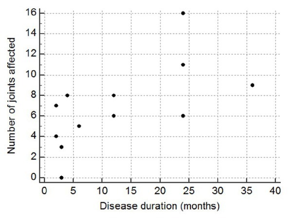
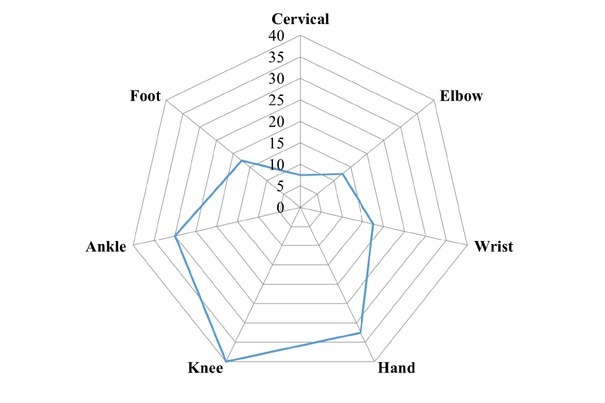
Results: The mean age of children was (8.05 ± 3.20) years of which 57.5% was male and the disease duration associated with the deformities in JIA. The prevalent subtypes of JIA were Oligoarticular JIA (oligoJIA)(40%), followed by polyarticular JIA (pJIA) (35%) and systemic-onset JIA (sJIA) (12.5%). The commonly involved joint were knee (40%), followed by small joint of hand (32.5%), ankle (30%), wrist and foot (17.5% each), elbow (12.5%) and cervical joint (7.5%). In pJIA, duration of disease significantly (P = 0.017) associated with the number of affected joints. Mostly, wrist, knee and ankle deformities were observed in oligoJIA, pJIA and sJIA. The angular deviation (mean ± SD) of right and left knee were (2° ± 4.16°) and (1.87° ± 5.12°) in oligoJIA, (13.36° ± 17.03°) and (12.5° ± 15.08°) in pJIA and (3° ± 6.71°) and (2.4° ± 5.37°) in sJIA. Right ankle angular deviation were (2.62° ± 5.06), (5.43° ± 8.21°) and 4° ± 8.94° respectively in oligoJIA, pJIA and sJIA. The angular deviation of right and left wrist were (1.25° ± 3.41°) and (0.94° ± 3.75°) in oligoJIA, (4.07° ± 8.93°) and (4.14° ± 9.36°) in pJIA and (2.45° ± 5.37°) and (2° ± 4.47°) in sJIA.
Conclusion: This study is the first study from India to quantify the angular deviation of deformed joints in JIA. Angular deviation could serve as a valuable parameter for monitoring disease progression across various JIA subtypes.

 求助内容:
求助内容: 应助结果提醒方式:


