Débora Costa Ruiz, Rocharles Cavalcante Fontenele, Amanda Farias-Gomes, Matheus L Oliveira, Deborah Queiroz Freitas, Francisco Haiter-Neto
{"title":"壁挂式和手持x光设备拍摄的x光片质量比较。","authors":"Débora Costa Ruiz, Rocharles Cavalcante Fontenele, Amanda Farias-Gomes, Matheus L Oliveira, Deborah Queiroz Freitas, Francisco Haiter-Neto","doi":"10.5624/isd.20240112","DOIUrl":null,"url":null,"abstract":"<p><strong>Purpose: </strong>This study was conducted to compare the objective image quality of radiographs acquired with a handheld X-ray device to those obtained with a wall-mounted X-ray device.</p><p><strong>Materials and methods: </strong>Brightness, noise, uniformity, and contrast were evaluated. To assess the first 3 parameters, radiographs of an acrylic block were acquired with an unused photostimulable phosphor (PSP) plate from the VistaScan system (Mini Easy, Dürr Dental, Bietigheim-Bissingen, Germany). Initially, 6 radiographs were taken with a Focus X-ray wall-mounted device (Instrumentarium, Tuusula, Finland) operating at 60 kVp, 7 mA, and 0.125 s. Another 6 radiographs were captured using an Eagle handheld X-ray device (Alliage, São Paulo, Brazil) at 60 kVp, 2.5 mA, and 0.35 s. The means and standard deviations of the gray values for all radiographs were calculated using ImageJ (National Institutes of Health, Bethesda, MD, USA). For contrast assessment, radiographs of an aluminum step wedge were obtained using the same PSP plate, X-ray devices, and acquisition parameters. The percentage of contrast variation was determined. The impacts of the devices on image quality were compared using the Student <i>t</i>-test, with a significance level of 5% (<i>P</i><0.05).</p><p><strong>Results: </strong>Compared with the wall-mounted device, the handheld device produced radiographs with higher brightness and noise, as indicated by mean values of 6.57 (0.49) and 3.49 (0.02), respectively. Furthermore, it demonstrated lower uniformity and contrast, with respective means of 3.75 (0.02) and 35.48 (0.09) (<i>P</i><0.05).</p><p><strong>Conclusion: </strong>Radiographs obtained using a handheld X-ray device exhibit lower theoretical image quality than those acquired with a wall-mounted device.</p>","PeriodicalId":51714,"journal":{"name":"Imaging Science in Dentistry","volume":"55 1","pages":"22-27"},"PeriodicalIF":2.1000,"publicationDate":"2025-03-01","publicationTypes":"Journal Article","fieldsOfStudy":null,"isOpenAccess":false,"openAccessPdf":"https://www.ncbi.nlm.nih.gov/pmc/articles/PMC11966020/pdf/","citationCount":"0","resultStr":"{\"title\":\"Comparison of objective radiograph quality between radiographs obtained with wall-mounted and handheld X-ray devices.\",\"authors\":\"Débora Costa Ruiz, Rocharles Cavalcante Fontenele, Amanda Farias-Gomes, Matheus L Oliveira, Deborah Queiroz Freitas, Francisco Haiter-Neto\",\"doi\":\"10.5624/isd.20240112\",\"DOIUrl\":null,\"url\":null,\"abstract\":\"<p><strong>Purpose: </strong>This study was conducted to compare the objective image quality of radiographs acquired with a handheld X-ray device to those obtained with a wall-mounted X-ray device.</p><p><strong>Materials and methods: </strong>Brightness, noise, uniformity, and contrast were evaluated. To assess the first 3 parameters, radiographs of an acrylic block were acquired with an unused photostimulable phosphor (PSP) plate from the VistaScan system (Mini Easy, Dürr Dental, Bietigheim-Bissingen, Germany). Initially, 6 radiographs were taken with a Focus X-ray wall-mounted device (Instrumentarium, Tuusula, Finland) operating at 60 kVp, 7 mA, and 0.125 s. Another 6 radiographs were captured using an Eagle handheld X-ray device (Alliage, São Paulo, Brazil) at 60 kVp, 2.5 mA, and 0.35 s. The means and standard deviations of the gray values for all radiographs were calculated using ImageJ (National Institutes of Health, Bethesda, MD, USA). For contrast assessment, radiographs of an aluminum step wedge were obtained using the same PSP plate, X-ray devices, and acquisition parameters. The percentage of contrast variation was determined. The impacts of the devices on image quality were compared using the Student <i>t</i>-test, with a significance level of 5% (<i>P</i><0.05).</p><p><strong>Results: </strong>Compared with the wall-mounted device, the handheld device produced radiographs with higher brightness and noise, as indicated by mean values of 6.57 (0.49) and 3.49 (0.02), respectively. Furthermore, it demonstrated lower uniformity and contrast, with respective means of 3.75 (0.02) and 35.48 (0.09) (<i>P</i><0.05).</p><p><strong>Conclusion: </strong>Radiographs obtained using a handheld X-ray device exhibit lower theoretical image quality than those acquired with a wall-mounted device.</p>\",\"PeriodicalId\":51714,\"journal\":{\"name\":\"Imaging Science in Dentistry\",\"volume\":\"55 1\",\"pages\":\"22-27\"},\"PeriodicalIF\":2.1000,\"publicationDate\":\"2025-03-01\",\"publicationTypes\":\"Journal Article\",\"fieldsOfStudy\":null,\"isOpenAccess\":false,\"openAccessPdf\":\"https://www.ncbi.nlm.nih.gov/pmc/articles/PMC11966020/pdf/\",\"citationCount\":\"0\",\"resultStr\":null,\"platform\":\"Semanticscholar\",\"paperid\":null,\"PeriodicalName\":\"Imaging Science in Dentistry\",\"FirstCategoryId\":\"1085\",\"ListUrlMain\":\"https://doi.org/10.5624/isd.20240112\",\"RegionNum\":0,\"RegionCategory\":null,\"ArticlePicture\":[],\"TitleCN\":null,\"AbstractTextCN\":null,\"PMCID\":null,\"EPubDate\":\"2024/12/6 0:00:00\",\"PubModel\":\"Epub\",\"JCR\":\"Q3\",\"JCRName\":\"DENTISTRY, ORAL SURGERY & MEDICINE\",\"Score\":null,\"Total\":0}","platform":"Semanticscholar","paperid":null,"PeriodicalName":"Imaging Science in Dentistry","FirstCategoryId":"1085","ListUrlMain":"https://doi.org/10.5624/isd.20240112","RegionNum":0,"RegionCategory":null,"ArticlePicture":[],"TitleCN":null,"AbstractTextCN":null,"PMCID":null,"EPubDate":"2024/12/6 0:00:00","PubModel":"Epub","JCR":"Q3","JCRName":"DENTISTRY, ORAL SURGERY & MEDICINE","Score":null,"Total":0}
引用次数: 0
摘要
目的:本研究比较了手持x光机和壁挂式x光机拍摄的x光片的客观图像质量。材料和方法:对亮度、噪声、均匀性和对比度进行评价。为了评估前3个参数,使用来自VistaScan系统(Mini Easy, d rr Dental, bietiheim - bissingen,德国)的未使用的光刺激荧光粉(PSP)板获取丙烯酸块的x线片。最初,使用Focus x射线壁挂式设备(instrumarium, Tuusula, Finland)在60 kVp, 7 mA, 0.125 s下拍摄6张x射线片。另外6张x光片使用Eagle手持式x光设备(Alliage, s o Paulo, Brazil)在60 kVp, 2.5 mA, 0.35 s下拍摄。使用ImageJ (National Institutes of Health, Bethesda, MD, USA)计算所有x线片灰度值的均值和标准差。为了进行对比评估,使用相同的PSP板、x射线设备和采集参数获得了铝阶梯楔的x线片。测定对比变化的百分比。使用Student t检验比较设备对图像质量的影响,显著性水平为5%(结果:与壁挂式设备相比,手持设备产生的x光片亮度和噪声更高,平均值分别为6.57(0.49)和3.49(0.02)。此外,它的均匀性和对比度较低,平均值分别为3.75(0.02)和35.48(0.09)。(结论:使用手持x射线设备获得的x射线片的理论图像质量低于使用壁挂式设备获得的x射线片。
Comparison of objective radiograph quality between radiographs obtained with wall-mounted and handheld X-ray devices.
Purpose: This study was conducted to compare the objective image quality of radiographs acquired with a handheld X-ray device to those obtained with a wall-mounted X-ray device.
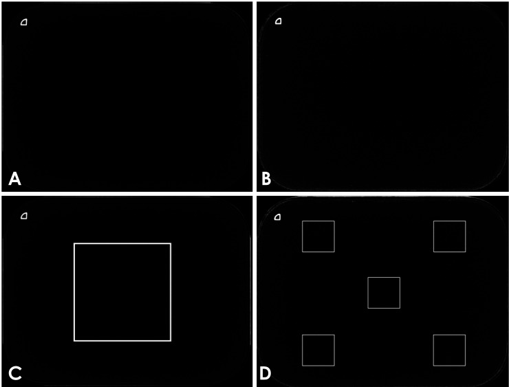
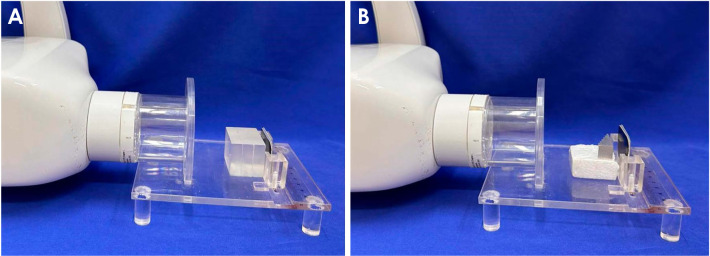
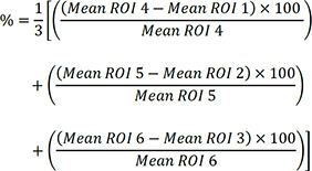
Materials and methods: Brightness, noise, uniformity, and contrast were evaluated. To assess the first 3 parameters, radiographs of an acrylic block were acquired with an unused photostimulable phosphor (PSP) plate from the VistaScan system (Mini Easy, Dürr Dental, Bietigheim-Bissingen, Germany). Initially, 6 radiographs were taken with a Focus X-ray wall-mounted device (Instrumentarium, Tuusula, Finland) operating at 60 kVp, 7 mA, and 0.125 s. Another 6 radiographs were captured using an Eagle handheld X-ray device (Alliage, São Paulo, Brazil) at 60 kVp, 2.5 mA, and 0.35 s. The means and standard deviations of the gray values for all radiographs were calculated using ImageJ (National Institutes of Health, Bethesda, MD, USA). For contrast assessment, radiographs of an aluminum step wedge were obtained using the same PSP plate, X-ray devices, and acquisition parameters. The percentage of contrast variation was determined. The impacts of the devices on image quality were compared using the Student t-test, with a significance level of 5% (P<0.05).
Results: Compared with the wall-mounted device, the handheld device produced radiographs with higher brightness and noise, as indicated by mean values of 6.57 (0.49) and 3.49 (0.02), respectively. Furthermore, it demonstrated lower uniformity and contrast, with respective means of 3.75 (0.02) and 35.48 (0.09) (P<0.05).
Conclusion: Radiographs obtained using a handheld X-ray device exhibit lower theoretical image quality than those acquired with a wall-mounted device.

 求助内容:
求助内容: 应助结果提醒方式:


