Baptiste Lodé, Burhan Rashid Hussein, Cédric Meurée, Ricky Walsh, Malo Gaubert, Nicolas Lassalle, Guilhem Courbon, Agathe Martin, Jeanne Le Bars, Françoise Durand-Dubief, Bertrand Bourre, Adil Maarouf, Olivier Outteryck, Clément Mehier, Alexandre Poulin, Camille Cathelineau, Jeremy Hong, Guillaume Criton, Sophie Motillon-Alonso, Augustin Lecler, Frédérique Charbonneau, Loïc Duron, Alexandre Bani-Sadr, Céline Delpierre, Jean-Christophe Ferré, Gilles Edan, François Cotton, Romain Casey, Francesca Galassi, Benoit Combès, Anne Kerbrat
{"title":"评估一种深度学习分割工具,以帮助从多发性硬化症患者的T2和STIR联合采集中检测脊髓病变。","authors":"Baptiste Lodé, Burhan Rashid Hussein, Cédric Meurée, Ricky Walsh, Malo Gaubert, Nicolas Lassalle, Guilhem Courbon, Agathe Martin, Jeanne Le Bars, Françoise Durand-Dubief, Bertrand Bourre, Adil Maarouf, Olivier Outteryck, Clément Mehier, Alexandre Poulin, Camille Cathelineau, Jeremy Hong, Guillaume Criton, Sophie Motillon-Alonso, Augustin Lecler, Frédérique Charbonneau, Loïc Duron, Alexandre Bani-Sadr, Céline Delpierre, Jean-Christophe Ferré, Gilles Edan, François Cotton, Romain Casey, Francesca Galassi, Benoit Combès, Anne Kerbrat","doi":"10.1007/s00330-025-11541-0","DOIUrl":null,"url":null,"abstract":"<p><strong>Objective: </strong>To develop a deep learning (DL) model for the detection of spinal cord (SC) multiple sclerosis (MS) lesions from both sagittal T2 and short tau inversion recovery (STIR) sequences and to investigate whether such a model could improve the performance of clinicians in detecting SC lesions.</p><p><strong>Materials and methods: </strong>A DL tool was developed based on SC sagittal T2 and STIR acquisitions from the imaging database of the French MS registry (OFSEP), including retrospective data from 40 different scanners. A multi-reader study based on retrospective data was performed between December 2023 and June 2024 to compare the performance of 20 clinicians in interpreting upper and lower SC acquisitions with and without the use of the tool. A ground truth was established by three experts. Sensitivity, precision, and inter-reader variability were evaluated.</p><p><strong>Results: </strong>We included 50 patients (39 females, median age: 41 years [range: 15-67]) with SC MRI acquired between February 2017 and December 2022. When reading with the tool, the clinicians' mean sensitivity to detect SC lesions improved (from 74.3% [95% CI = 67.8-80.6%] to 79.2% [95% CI: 73.5-85.0%]; p < 0.0001), with no evidence of difference in the mean precision: (69.0% [95% CI: 62.8-75.2%] vs 70.1% [95% CI: 64.3-75.9%]; p = 0.08). Inter-reader variability in lesion detection was slightly improved with the tool (Light's kappa = 0.55 vs 0.60), but without statistical difference (p = 0.056).</p><p><strong>Conclusion: </strong>The use of an automatic tool can help clinicians detect SC lesions in pwMS by increasing their sensitivity.</p><p><strong>Key points: </strong>Question No tool to help detect MS SC lesions is used in clinical practice despite their frequency and prognostic value. Findings This DL-based tool led to improvement in clinicians' sensitivity in detecting SC lesions from both sagittal T2 and STIR sequences, without decreasing precision. Clinical relevance Our study indicated the potential of a DL-based tool to assist clinicians in the challenging task of detecting SC lesions in people with MS on a combination of sequences commonly acquired in clinical practice.</p>","PeriodicalId":12076,"journal":{"name":"European Radiology","volume":" ","pages":"5954-5964"},"PeriodicalIF":4.7000,"publicationDate":"2025-10-01","publicationTypes":"Journal Article","fieldsOfStudy":null,"isOpenAccess":false,"openAccessPdf":"https://www.ncbi.nlm.nih.gov/pmc/articles/PMC12417267/pdf/","citationCount":"0","resultStr":"{\"title\":\"Evaluation of a deep learning segmentation tool to help detect spinal cord lesions from combined T2 and STIR acquisitions in people with multiple sclerosis.\",\"authors\":\"Baptiste Lodé, Burhan Rashid Hussein, Cédric Meurée, Ricky Walsh, Malo Gaubert, Nicolas Lassalle, Guilhem Courbon, Agathe Martin, Jeanne Le Bars, Françoise Durand-Dubief, Bertrand Bourre, Adil Maarouf, Olivier Outteryck, Clément Mehier, Alexandre Poulin, Camille Cathelineau, Jeremy Hong, Guillaume Criton, Sophie Motillon-Alonso, Augustin Lecler, Frédérique Charbonneau, Loïc Duron, Alexandre Bani-Sadr, Céline Delpierre, Jean-Christophe Ferré, Gilles Edan, François Cotton, Romain Casey, Francesca Galassi, Benoit Combès, Anne Kerbrat\",\"doi\":\"10.1007/s00330-025-11541-0\",\"DOIUrl\":null,\"url\":null,\"abstract\":\"<p><strong>Objective: </strong>To develop a deep learning (DL) model for the detection of spinal cord (SC) multiple sclerosis (MS) lesions from both sagittal T2 and short tau inversion recovery (STIR) sequences and to investigate whether such a model could improve the performance of clinicians in detecting SC lesions.</p><p><strong>Materials and methods: </strong>A DL tool was developed based on SC sagittal T2 and STIR acquisitions from the imaging database of the French MS registry (OFSEP), including retrospective data from 40 different scanners. A multi-reader study based on retrospective data was performed between December 2023 and June 2024 to compare the performance of 20 clinicians in interpreting upper and lower SC acquisitions with and without the use of the tool. A ground truth was established by three experts. Sensitivity, precision, and inter-reader variability were evaluated.</p><p><strong>Results: </strong>We included 50 patients (39 females, median age: 41 years [range: 15-67]) with SC MRI acquired between February 2017 and December 2022. When reading with the tool, the clinicians' mean sensitivity to detect SC lesions improved (from 74.3% [95% CI = 67.8-80.6%] to 79.2% [95% CI: 73.5-85.0%]; p < 0.0001), with no evidence of difference in the mean precision: (69.0% [95% CI: 62.8-75.2%] vs 70.1% [95% CI: 64.3-75.9%]; p = 0.08). Inter-reader variability in lesion detection was slightly improved with the tool (Light's kappa = 0.55 vs 0.60), but without statistical difference (p = 0.056).</p><p><strong>Conclusion: </strong>The use of an automatic tool can help clinicians detect SC lesions in pwMS by increasing their sensitivity.</p><p><strong>Key points: </strong>Question No tool to help detect MS SC lesions is used in clinical practice despite their frequency and prognostic value. Findings This DL-based tool led to improvement in clinicians' sensitivity in detecting SC lesions from both sagittal T2 and STIR sequences, without decreasing precision. Clinical relevance Our study indicated the potential of a DL-based tool to assist clinicians in the challenging task of detecting SC lesions in people with MS on a combination of sequences commonly acquired in clinical practice.</p>\",\"PeriodicalId\":12076,\"journal\":{\"name\":\"European Radiology\",\"volume\":\" \",\"pages\":\"5954-5964\"},\"PeriodicalIF\":4.7000,\"publicationDate\":\"2025-10-01\",\"publicationTypes\":\"Journal Article\",\"fieldsOfStudy\":null,\"isOpenAccess\":false,\"openAccessPdf\":\"https://www.ncbi.nlm.nih.gov/pmc/articles/PMC12417267/pdf/\",\"citationCount\":\"0\",\"resultStr\":null,\"platform\":\"Semanticscholar\",\"paperid\":null,\"PeriodicalName\":\"European Radiology\",\"FirstCategoryId\":\"3\",\"ListUrlMain\":\"https://doi.org/10.1007/s00330-025-11541-0\",\"RegionNum\":2,\"RegionCategory\":\"医学\",\"ArticlePicture\":[],\"TitleCN\":null,\"AbstractTextCN\":null,\"PMCID\":null,\"EPubDate\":\"2025/4/4 0:00:00\",\"PubModel\":\"Epub\",\"JCR\":\"Q1\",\"JCRName\":\"RADIOLOGY, NUCLEAR MEDICINE & MEDICAL IMAGING\",\"Score\":null,\"Total\":0}","platform":"Semanticscholar","paperid":null,"PeriodicalName":"European Radiology","FirstCategoryId":"3","ListUrlMain":"https://doi.org/10.1007/s00330-025-11541-0","RegionNum":2,"RegionCategory":"医学","ArticlePicture":[],"TitleCN":null,"AbstractTextCN":null,"PMCID":null,"EPubDate":"2025/4/4 0:00:00","PubModel":"Epub","JCR":"Q1","JCRName":"RADIOLOGY, NUCLEAR MEDICINE & MEDICAL IMAGING","Score":null,"Total":0}
Evaluation of a deep learning segmentation tool to help detect spinal cord lesions from combined T2 and STIR acquisitions in people with multiple sclerosis.
Objective: To develop a deep learning (DL) model for the detection of spinal cord (SC) multiple sclerosis (MS) lesions from both sagittal T2 and short tau inversion recovery (STIR) sequences and to investigate whether such a model could improve the performance of clinicians in detecting SC lesions.
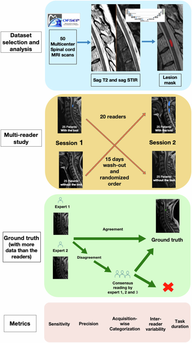
Materials and methods: A DL tool was developed based on SC sagittal T2 and STIR acquisitions from the imaging database of the French MS registry (OFSEP), including retrospective data from 40 different scanners. A multi-reader study based on retrospective data was performed between December 2023 and June 2024 to compare the performance of 20 clinicians in interpreting upper and lower SC acquisitions with and without the use of the tool. A ground truth was established by three experts. Sensitivity, precision, and inter-reader variability were evaluated.
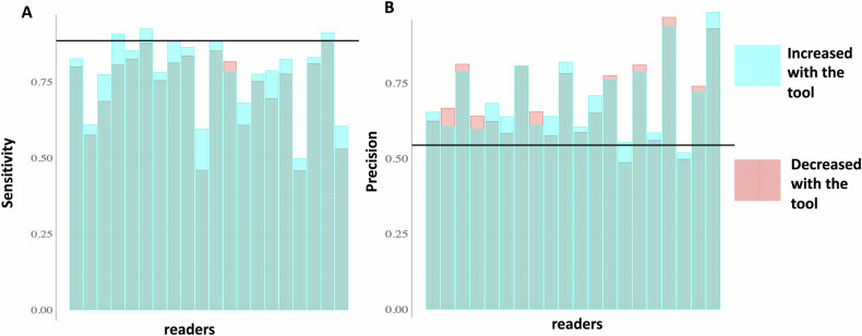
Results: We included 50 patients (39 females, median age: 41 years [range: 15-67]) with SC MRI acquired between February 2017 and December 2022. When reading with the tool, the clinicians' mean sensitivity to detect SC lesions improved (from 74.3% [95% CI = 67.8-80.6%] to 79.2% [95% CI: 73.5-85.0%]; p < 0.0001), with no evidence of difference in the mean precision: (69.0% [95% CI: 62.8-75.2%] vs 70.1% [95% CI: 64.3-75.9%]; p = 0.08). Inter-reader variability in lesion detection was slightly improved with the tool (Light's kappa = 0.55 vs 0.60), but without statistical difference (p = 0.056).
Conclusion: The use of an automatic tool can help clinicians detect SC lesions in pwMS by increasing their sensitivity.
Key points: Question No tool to help detect MS SC lesions is used in clinical practice despite their frequency and prognostic value. Findings This DL-based tool led to improvement in clinicians' sensitivity in detecting SC lesions from both sagittal T2 and STIR sequences, without decreasing precision. Clinical relevance Our study indicated the potential of a DL-based tool to assist clinicians in the challenging task of detecting SC lesions in people with MS on a combination of sequences commonly acquired in clinical practice.
期刊介绍:
European Radiology (ER) continuously updates scientific knowledge in radiology by publication of strong original articles and state-of-the-art reviews written by leading radiologists. A well balanced combination of review articles, original papers, short communications from European radiological congresses and information on society matters makes ER an indispensable source for current information in this field.
This is the Journal of the European Society of Radiology, and the official journal of a number of societies.
From 2004-2008 supplements to European Radiology were published under its companion, European Radiology Supplements, ISSN 1613-3749.

 求助内容:
求助内容: 应助结果提醒方式:


