Ramya Gnanaraj, Andres Lisker-Cervantes, Jennifer Patnaik, Vivian Rajeswaren, Nihaal Mehta, William Gange, Anne M Lynch, Alan Palestine, Marc Mathias, Niranjan Manoharan, Naresh Mandava, Talisa E de Carlo Forest
{"title":"从中晚期年龄相关性黄斑变性(AMD)进展的多模态成像生物标志物:一项来自科罗拉多大学AMD注册中心的10年前瞻性纵向队列研究。","authors":"Ramya Gnanaraj, Andres Lisker-Cervantes, Jennifer Patnaik, Vivian Rajeswaren, Nihaal Mehta, William Gange, Anne M Lynch, Alan Palestine, Marc Mathias, Niranjan Manoharan, Naresh Mandava, Talisa E de Carlo Forest","doi":"10.1136/bmjophth-2024-002112","DOIUrl":null,"url":null,"abstract":"<p><strong>Objective: </strong>To evaluate multimodal imaging (MMI) biomarkers for predicting progression from intermediate to advanced age-related macular degeneration (AMD).</p><p><strong>Methods and analysis: </strong>This prospective longitudinal cohort study included patients with intermediate AMD (iAMD) enrolled in the University of Colorado AMD registry between July 2014 and August 2023, with follow-up through February 2024. At enrolment, patients' medical histories and MMI were collected. Baseline and follow-up imaging were reviewed for progression to geographic atrophy (GA) and neovascular AMD (nAMD). Univariate and multivariable Cox proportional hazard modelling with competing risks to determine HRs for progression.</p><p><strong>Results: </strong>A total of 367 patients (733 eyes) with iAMD were included in the study, with a median follow-up of 27.8 months. During this period, 100 eyes progressed to GA, 58 to nAMD. Adjusted for age, BMI and hypertension, progression to nAMD was significantly associated with soft drusen (HR 5.31, 95% CI 1.95 to 14.4, p=0.001), pigmentary changes (HR 2.74, 95% CI 1.52 to 4.92, p=0.0008) on colour fundus photography (CFP) and subretinal hyper-reflective material (SHRM) (HR 3.36, 95% CI 1.88 to 6.02, p<0.0001) and intraretinal hyper-reflective foci (IHRF) (HR 3.12, 95% CI 1.74 to 5.57, p=0.0001) on optical coherence tomography (OCT). Adjusted for age, progression to GA was predicted by soft drusen (HR 1.90, 95% CI 1.11 to 3.27, p=0.020), drusenoid pigment epithelial detachment (PED) (HR 5.51, 95% CI 2.49 to 12.2, p<0.0001), avascular non-drusenoid PED (HR 6.59, 95% CI 1.54 to 28.1, p=0.011), pigmentary changes (HR 4.44, 95% CI 2.84 to 6.96, p<0.0001) on CFP and nnSRF (HR 6.41, 95% CI 1.39 to 29.6, p=0.017), SHRM (HR 2.55, 95% CI 1.45 to 4.49, p=0.001), drusenoid PED (HR 2.25, 95% CI 1.43 to 3.55, p=0.0005), avascular non-drusenoid PED (HR 4.67, 95% CI 2.45 to 8.92, p<0.0001), IHRF (HR 6.27, 95% CI 3.89 to 10.1, p<0.0001) and incomplete retinal pigment epithelium and outer retinal atrophy (HR 9.42, 95% CI 5.82 to 15.2, p<0.0001) on OCT (table 3).</p><p><strong>Conclusions: </strong>Key imaging biomarkers associated with the progression were identified, which may offer prognostic information for providers. However, the study is limited by its predominantly Caucasian population and single-centre design, which may affect the generalisability of certain biomarkers.</p>","PeriodicalId":9286,"journal":{"name":"BMJ Open Ophthalmology","volume":"10 1","pages":""},"PeriodicalIF":2.2000,"publicationDate":"2025-04-05","publicationTypes":"Journal Article","fieldsOfStudy":null,"isOpenAccess":false,"openAccessPdf":"https://www.ncbi.nlm.nih.gov/pmc/articles/PMC11973788/pdf/","citationCount":"0","resultStr":"{\"title\":\"Multimodal imaging biomarkers for progression from intermediate to advanced age-related macular degeneration (AMD): a 10-year prospective longitudinal cohort study from the University of Colorado AMD registry.\",\"authors\":\"Ramya Gnanaraj, Andres Lisker-Cervantes, Jennifer Patnaik, Vivian Rajeswaren, Nihaal Mehta, William Gange, Anne M Lynch, Alan Palestine, Marc Mathias, Niranjan Manoharan, Naresh Mandava, Talisa E de Carlo Forest\",\"doi\":\"10.1136/bmjophth-2024-002112\",\"DOIUrl\":null,\"url\":null,\"abstract\":\"<p><strong>Objective: </strong>To evaluate multimodal imaging (MMI) biomarkers for predicting progression from intermediate to advanced age-related macular degeneration (AMD).</p><p><strong>Methods and analysis: </strong>This prospective longitudinal cohort study included patients with intermediate AMD (iAMD) enrolled in the University of Colorado AMD registry between July 2014 and August 2023, with follow-up through February 2024. At enrolment, patients' medical histories and MMI were collected. Baseline and follow-up imaging were reviewed for progression to geographic atrophy (GA) and neovascular AMD (nAMD). Univariate and multivariable Cox proportional hazard modelling with competing risks to determine HRs for progression.</p><p><strong>Results: </strong>A total of 367 patients (733 eyes) with iAMD were included in the study, with a median follow-up of 27.8 months. During this period, 100 eyes progressed to GA, 58 to nAMD. Adjusted for age, BMI and hypertension, progression to nAMD was significantly associated with soft drusen (HR 5.31, 95% CI 1.95 to 14.4, p=0.001), pigmentary changes (HR 2.74, 95% CI 1.52 to 4.92, p=0.0008) on colour fundus photography (CFP) and subretinal hyper-reflective material (SHRM) (HR 3.36, 95% CI 1.88 to 6.02, p<0.0001) and intraretinal hyper-reflective foci (IHRF) (HR 3.12, 95% CI 1.74 to 5.57, p=0.0001) on optical coherence tomography (OCT). Adjusted for age, progression to GA was predicted by soft drusen (HR 1.90, 95% CI 1.11 to 3.27, p=0.020), drusenoid pigment epithelial detachment (PED) (HR 5.51, 95% CI 2.49 to 12.2, p<0.0001), avascular non-drusenoid PED (HR 6.59, 95% CI 1.54 to 28.1, p=0.011), pigmentary changes (HR 4.44, 95% CI 2.84 to 6.96, p<0.0001) on CFP and nnSRF (HR 6.41, 95% CI 1.39 to 29.6, p=0.017), SHRM (HR 2.55, 95% CI 1.45 to 4.49, p=0.001), drusenoid PED (HR 2.25, 95% CI 1.43 to 3.55, p=0.0005), avascular non-drusenoid PED (HR 4.67, 95% CI 2.45 to 8.92, p<0.0001), IHRF (HR 6.27, 95% CI 3.89 to 10.1, p<0.0001) and incomplete retinal pigment epithelium and outer retinal atrophy (HR 9.42, 95% CI 5.82 to 15.2, p<0.0001) on OCT (table 3).</p><p><strong>Conclusions: </strong>Key imaging biomarkers associated with the progression were identified, which may offer prognostic information for providers. However, the study is limited by its predominantly Caucasian population and single-centre design, which may affect the generalisability of certain biomarkers.</p>\",\"PeriodicalId\":9286,\"journal\":{\"name\":\"BMJ Open Ophthalmology\",\"volume\":\"10 1\",\"pages\":\"\"},\"PeriodicalIF\":2.2000,\"publicationDate\":\"2025-04-05\",\"publicationTypes\":\"Journal Article\",\"fieldsOfStudy\":null,\"isOpenAccess\":false,\"openAccessPdf\":\"https://www.ncbi.nlm.nih.gov/pmc/articles/PMC11973788/pdf/\",\"citationCount\":\"0\",\"resultStr\":null,\"platform\":\"Semanticscholar\",\"paperid\":null,\"PeriodicalName\":\"BMJ Open Ophthalmology\",\"FirstCategoryId\":\"1085\",\"ListUrlMain\":\"https://doi.org/10.1136/bmjophth-2024-002112\",\"RegionNum\":0,\"RegionCategory\":null,\"ArticlePicture\":[],\"TitleCN\":null,\"AbstractTextCN\":null,\"PMCID\":null,\"EPubDate\":\"\",\"PubModel\":\"\",\"JCR\":\"Q2\",\"JCRName\":\"OPHTHALMOLOGY\",\"Score\":null,\"Total\":0}","platform":"Semanticscholar","paperid":null,"PeriodicalName":"BMJ Open Ophthalmology","FirstCategoryId":"1085","ListUrlMain":"https://doi.org/10.1136/bmjophth-2024-002112","RegionNum":0,"RegionCategory":null,"ArticlePicture":[],"TitleCN":null,"AbstractTextCN":null,"PMCID":null,"EPubDate":"","PubModel":"","JCR":"Q2","JCRName":"OPHTHALMOLOGY","Score":null,"Total":0}
引用次数: 0
摘要
目的:评价多模态成像(MMI)生物标志物预测中晚期老年性黄斑变性(AMD)的进展。方法和分析:这项前瞻性纵向队列研究纳入了2014年7月至2023年8月在科罗拉多大学AMD注册中心注册的中度AMD (iAMD)患者,随访至2024年2月。在入组时,收集患者的病史和MMI。基线和随访影像学检查进展为地理萎缩(GA)和新生血管性AMD (nAMD)。具有竞争风险的单变量和多变量Cox比例风险模型,以确定进展的hr。结果:共有367例iAMD患者(733只眼)纳入研究,中位随访时间为27.8个月。在此期间,100只眼睛进展为GA, 58只进展为nAMD。经年龄、BMI和高血压校正后,进展为nAMD与眼底彩色摄影(CFP)的软性肾结石(HR 5.31, 95% CI 1.95 ~ 14.4, p=0.001)、色素改变(HR 2.74, 95% CI 1.52 ~ 4.92, p=0.0008)和视网膜下高反射物质(HR 3.36, 95% CI 1.88 ~ 6.02, p)显著相关。结论:确定了与进展相关的关键成像生物标志物,这可能为提供者提供预后信息。然而,该研究受限于其主要的高加索人群和单中心设计,这可能会影响某些生物标志物的普遍性。
Multimodal imaging biomarkers for progression from intermediate to advanced age-related macular degeneration (AMD): a 10-year prospective longitudinal cohort study from the University of Colorado AMD registry.
Objective: To evaluate multimodal imaging (MMI) biomarkers for predicting progression from intermediate to advanced age-related macular degeneration (AMD).
Methods and analysis: This prospective longitudinal cohort study included patients with intermediate AMD (iAMD) enrolled in the University of Colorado AMD registry between July 2014 and August 2023, with follow-up through February 2024. At enrolment, patients' medical histories and MMI were collected. Baseline and follow-up imaging were reviewed for progression to geographic atrophy (GA) and neovascular AMD (nAMD). Univariate and multivariable Cox proportional hazard modelling with competing risks to determine HRs for progression.
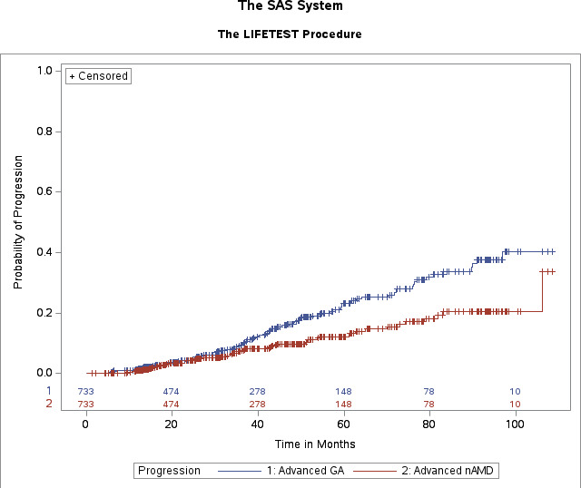
Results: A total of 367 patients (733 eyes) with iAMD were included in the study, with a median follow-up of 27.8 months. During this period, 100 eyes progressed to GA, 58 to nAMD. Adjusted for age, BMI and hypertension, progression to nAMD was significantly associated with soft drusen (HR 5.31, 95% CI 1.95 to 14.4, p=0.001), pigmentary changes (HR 2.74, 95% CI 1.52 to 4.92, p=0.0008) on colour fundus photography (CFP) and subretinal hyper-reflective material (SHRM) (HR 3.36, 95% CI 1.88 to 6.02, p<0.0001) and intraretinal hyper-reflective foci (IHRF) (HR 3.12, 95% CI 1.74 to 5.57, p=0.0001) on optical coherence tomography (OCT). Adjusted for age, progression to GA was predicted by soft drusen (HR 1.90, 95% CI 1.11 to 3.27, p=0.020), drusenoid pigment epithelial detachment (PED) (HR 5.51, 95% CI 2.49 to 12.2, p<0.0001), avascular non-drusenoid PED (HR 6.59, 95% CI 1.54 to 28.1, p=0.011), pigmentary changes (HR 4.44, 95% CI 2.84 to 6.96, p<0.0001) on CFP and nnSRF (HR 6.41, 95% CI 1.39 to 29.6, p=0.017), SHRM (HR 2.55, 95% CI 1.45 to 4.49, p=0.001), drusenoid PED (HR 2.25, 95% CI 1.43 to 3.55, p=0.0005), avascular non-drusenoid PED (HR 4.67, 95% CI 2.45 to 8.92, p<0.0001), IHRF (HR 6.27, 95% CI 3.89 to 10.1, p<0.0001) and incomplete retinal pigment epithelium and outer retinal atrophy (HR 9.42, 95% CI 5.82 to 15.2, p<0.0001) on OCT (table 3).
Conclusions: Key imaging biomarkers associated with the progression were identified, which may offer prognostic information for providers. However, the study is limited by its predominantly Caucasian population and single-centre design, which may affect the generalisability of certain biomarkers.

 求助内容:
求助内容: 应助结果提醒方式:


