Felicia Forseni Flodin, Sven Haller, Leo Poom, David Fällmar
{"title":"内侧颞叶萎缩的公开图像显示之间的一致性。","authors":"Felicia Forseni Flodin, Sven Haller, Leo Poom, David Fällmar","doi":"10.1007/s00330-025-11529-w","DOIUrl":null,"url":null,"abstract":"<p><p>The medial temporal lobe atrophy (MTA) score is used for visual assessment of MTA on radiological images in suspected neurodegenerative dementia. Although volumetric tools are available, many radiologists still use visual scoring and compare to reference images. Numerous such example images are found online on educational websites and in scientific articles. The aim of this study was to compare congruencies between MTA scores of publicly available sample images with normalized heights and areas of relevant brain structures, measured in the same images.</p><p><strong>Method: </strong>Systematic online searches yielded 148 individual sample images. The height and area of relevant brain structures were manually delineated, normalized, and compared with regard to the displayed MTA score.</p><p><strong>Results: </strong>The normalized heights and areas showed correlation with MTA but with considerable overlap between adjacent scores, especially when comparing heights. Also, displays of the MTA score were more consistent with the area of the temporal horn than with the hippocampal area.</p><p><strong>Conclusion: </strong>There is considerable overlap between adjacent scores in publicly available pictorial displays of the MTA grading system. Insufficient congruency leads to confusion and reduces inter-rater reliability. We also found that publicly available images are more consistent with temporal horn area than the hippocampus, which means that ventricular size may bias the grading. This can impede relevant differential diagnostics, especially regarding normal pressure hydrocephalus. Here, we present lectotype images selected specifically with regard to the hippocampal area.</p><p><strong>Key points: </strong>Question Overlap between publicly available example images of medial temporal atrophy causes confusion and limits reliability. Findings Available images are more consistent with ventricular dilatation than hippocampal atrophy; this article provides lectotype images selected specifically regarding the hippocampal area. Clinical relevance Visual assessment of medial temporal atrophy is used daily and worldwide in radiological examinations regarding suspected dementia. In clinical routine, many radiologists experience uncertainty, and hydrocephalus is often overlooked. This may be caused by insufficient congruency between educational sample images.</p>","PeriodicalId":12076,"journal":{"name":"European Radiology","volume":" ","pages":"5944-5953"},"PeriodicalIF":4.7000,"publicationDate":"2025-10-01","publicationTypes":"Journal Article","fieldsOfStudy":null,"isOpenAccess":false,"openAccessPdf":"https://www.ncbi.nlm.nih.gov/pmc/articles/PMC12417228/pdf/","citationCount":"0","resultStr":"{\"title\":\"Congruency between publicly available pictorial displays of medial temporal lobe atrophy.\",\"authors\":\"Felicia Forseni Flodin, Sven Haller, Leo Poom, David Fällmar\",\"doi\":\"10.1007/s00330-025-11529-w\",\"DOIUrl\":null,\"url\":null,\"abstract\":\"<p><p>The medial temporal lobe atrophy (MTA) score is used for visual assessment of MTA on radiological images in suspected neurodegenerative dementia. Although volumetric tools are available, many radiologists still use visual scoring and compare to reference images. Numerous such example images are found online on educational websites and in scientific articles. The aim of this study was to compare congruencies between MTA scores of publicly available sample images with normalized heights and areas of relevant brain structures, measured in the same images.</p><p><strong>Method: </strong>Systematic online searches yielded 148 individual sample images. The height and area of relevant brain structures were manually delineated, normalized, and compared with regard to the displayed MTA score.</p><p><strong>Results: </strong>The normalized heights and areas showed correlation with MTA but with considerable overlap between adjacent scores, especially when comparing heights. Also, displays of the MTA score were more consistent with the area of the temporal horn than with the hippocampal area.</p><p><strong>Conclusion: </strong>There is considerable overlap between adjacent scores in publicly available pictorial displays of the MTA grading system. Insufficient congruency leads to confusion and reduces inter-rater reliability. We also found that publicly available images are more consistent with temporal horn area than the hippocampus, which means that ventricular size may bias the grading. This can impede relevant differential diagnostics, especially regarding normal pressure hydrocephalus. Here, we present lectotype images selected specifically with regard to the hippocampal area.</p><p><strong>Key points: </strong>Question Overlap between publicly available example images of medial temporal atrophy causes confusion and limits reliability. Findings Available images are more consistent with ventricular dilatation than hippocampal atrophy; this article provides lectotype images selected specifically regarding the hippocampal area. Clinical relevance Visual assessment of medial temporal atrophy is used daily and worldwide in radiological examinations regarding suspected dementia. In clinical routine, many radiologists experience uncertainty, and hydrocephalus is often overlooked. This may be caused by insufficient congruency between educational sample images.</p>\",\"PeriodicalId\":12076,\"journal\":{\"name\":\"European Radiology\",\"volume\":\" \",\"pages\":\"5944-5953\"},\"PeriodicalIF\":4.7000,\"publicationDate\":\"2025-10-01\",\"publicationTypes\":\"Journal Article\",\"fieldsOfStudy\":null,\"isOpenAccess\":false,\"openAccessPdf\":\"https://www.ncbi.nlm.nih.gov/pmc/articles/PMC12417228/pdf/\",\"citationCount\":\"0\",\"resultStr\":null,\"platform\":\"Semanticscholar\",\"paperid\":null,\"PeriodicalName\":\"European Radiology\",\"FirstCategoryId\":\"3\",\"ListUrlMain\":\"https://doi.org/10.1007/s00330-025-11529-w\",\"RegionNum\":2,\"RegionCategory\":\"医学\",\"ArticlePicture\":[],\"TitleCN\":null,\"AbstractTextCN\":null,\"PMCID\":null,\"EPubDate\":\"2025/4/3 0:00:00\",\"PubModel\":\"Epub\",\"JCR\":\"Q1\",\"JCRName\":\"RADIOLOGY, NUCLEAR MEDICINE & MEDICAL IMAGING\",\"Score\":null,\"Total\":0}","platform":"Semanticscholar","paperid":null,"PeriodicalName":"European Radiology","FirstCategoryId":"3","ListUrlMain":"https://doi.org/10.1007/s00330-025-11529-w","RegionNum":2,"RegionCategory":"医学","ArticlePicture":[],"TitleCN":null,"AbstractTextCN":null,"PMCID":null,"EPubDate":"2025/4/3 0:00:00","PubModel":"Epub","JCR":"Q1","JCRName":"RADIOLOGY, NUCLEAR MEDICINE & MEDICAL IMAGING","Score":null,"Total":0}
Congruency between publicly available pictorial displays of medial temporal lobe atrophy.
The medial temporal lobe atrophy (MTA) score is used for visual assessment of MTA on radiological images in suspected neurodegenerative dementia. Although volumetric tools are available, many radiologists still use visual scoring and compare to reference images. Numerous such example images are found online on educational websites and in scientific articles. The aim of this study was to compare congruencies between MTA scores of publicly available sample images with normalized heights and areas of relevant brain structures, measured in the same images.
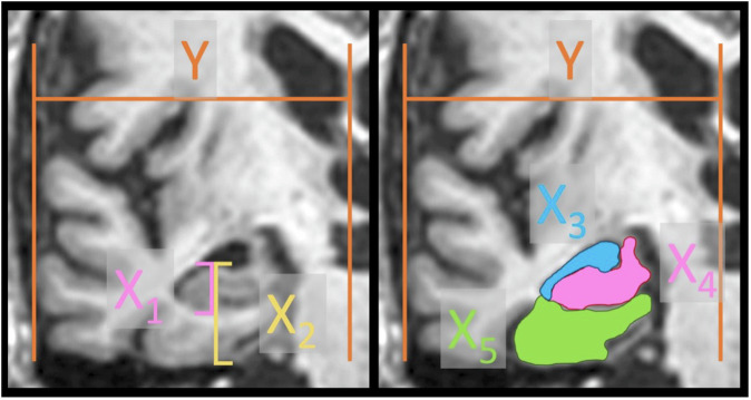
Method: Systematic online searches yielded 148 individual sample images. The height and area of relevant brain structures were manually delineated, normalized, and compared with regard to the displayed MTA score.
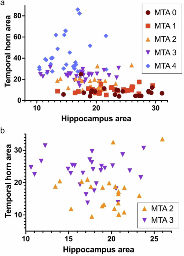
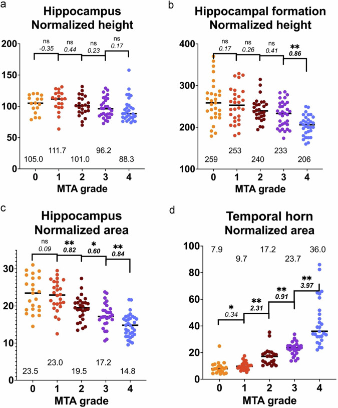
Results: The normalized heights and areas showed correlation with MTA but with considerable overlap between adjacent scores, especially when comparing heights. Also, displays of the MTA score were more consistent with the area of the temporal horn than with the hippocampal area.
Conclusion: There is considerable overlap between adjacent scores in publicly available pictorial displays of the MTA grading system. Insufficient congruency leads to confusion and reduces inter-rater reliability. We also found that publicly available images are more consistent with temporal horn area than the hippocampus, which means that ventricular size may bias the grading. This can impede relevant differential diagnostics, especially regarding normal pressure hydrocephalus. Here, we present lectotype images selected specifically with regard to the hippocampal area.
Key points: Question Overlap between publicly available example images of medial temporal atrophy causes confusion and limits reliability. Findings Available images are more consistent with ventricular dilatation than hippocampal atrophy; this article provides lectotype images selected specifically regarding the hippocampal area. Clinical relevance Visual assessment of medial temporal atrophy is used daily and worldwide in radiological examinations regarding suspected dementia. In clinical routine, many radiologists experience uncertainty, and hydrocephalus is often overlooked. This may be caused by insufficient congruency between educational sample images.
期刊介绍:
European Radiology (ER) continuously updates scientific knowledge in radiology by publication of strong original articles and state-of-the-art reviews written by leading radiologists. A well balanced combination of review articles, original papers, short communications from European radiological congresses and information on society matters makes ER an indispensable source for current information in this field.
This is the Journal of the European Society of Radiology, and the official journal of a number of societies.
From 2004-2008 supplements to European Radiology were published under its companion, European Radiology Supplements, ISSN 1613-3749.

 求助内容:
求助内容: 应助结果提醒方式:


