{"title":"缺血性脑卒中患者急性和亚急性期离子和细胞毒性水肿的动力学:23Na MRI和弥散MRI的互补","authors":"Maëva Cotinat, Noëlle Messaoudi, Emmanuelle Robinet, Laurent Suissa, Emilie Doche, Maxime Guye, Bertrand Audoin, Laurent Bensoussan, Jean-Philippe Ranjeva, Wafaa Zaaraoui","doi":"10.1002/nbm.70028","DOIUrl":null,"url":null,"abstract":"<p><p>Cerebral imaging is crucial in the diagnosis and treatment algorithm of acute stroke to determine salvageable brain tissue. While diffusion MRI is commonly used to define the ischemic core, it cannot reliably distinguish irreversibly damaged from salvageable tissue. We investigated the added value of <sup>23</sup>Na MRI to define irreversible necrotic tissue after a stroke. Fifteen patients with acute stroke involving medial cerebral artery occlusion were longitudinally explored with conventional and <sup>23</sup>Na MRI within 24 h, 70 h following stroke and at 3 months to characterize the necrotic area. Time-courses of sodium accumulations were observed within regions presenting with or spared by cytotoxic/ionic edema and converting or not to necrosis. Dynamics of sodium accumulations were very different across subjects. At the group level, time-courses of sodium signal in cytotoxic edema showed a non-linear increase with an upper asymptote of 59 ± 6%% relative to the contralateral hemisphere. Regions with a larger early increase in <sup>23</sup>Na signal (ionic edema) showed a non-linear accumulation during the first 70 h and were associated with subsequent necrosis at month 3. Some of the regions with no ionic edema during the first 70 h became necrotic at month 3, showing that pejorative pathophysiological processes could worsen after 70 h following attack. Final necrotic volume was well predicted by the cytotoxic volume (ADC decrease) during the first 24 h, and by the volume of ionic edema during the subacute period (25-70 h) following attack. The regions showing ionic edema showed a non-linear increase of <sup>23</sup>Na signal during the first 70 h, with larger sodium accumulations in regions converting to necrosis at month 3. It may be of interest to consider the role of ionic edema imaging in the 70 h after stroke and reperfusion, with a view to better understand stroke pathophysiology. Sodium MRI could add complementary information about the fate of cell necrosis within low ADC signal regions.</p>","PeriodicalId":19309,"journal":{"name":"NMR in Biomedicine","volume":"38 5","pages":"e70028"},"PeriodicalIF":2.7000,"publicationDate":"2025-05-01","publicationTypes":"Journal Article","fieldsOfStudy":null,"isOpenAccess":false,"openAccessPdf":"https://www.ncbi.nlm.nih.gov/pmc/articles/PMC11964797/pdf/","citationCount":"0","resultStr":"{\"title\":\"Dynamics of Ionic and Cytotoxic Edema During Acute and Subacute Stages of Patients With Ischemic Stroke: Complementarity of <sup>23</sup>Na MRI and Diffusion MRI.\",\"authors\":\"Maëva Cotinat, Noëlle Messaoudi, Emmanuelle Robinet, Laurent Suissa, Emilie Doche, Maxime Guye, Bertrand Audoin, Laurent Bensoussan, Jean-Philippe Ranjeva, Wafaa Zaaraoui\",\"doi\":\"10.1002/nbm.70028\",\"DOIUrl\":null,\"url\":null,\"abstract\":\"<p><p>Cerebral imaging is crucial in the diagnosis and treatment algorithm of acute stroke to determine salvageable brain tissue. While diffusion MRI is commonly used to define the ischemic core, it cannot reliably distinguish irreversibly damaged from salvageable tissue. We investigated the added value of <sup>23</sup>Na MRI to define irreversible necrotic tissue after a stroke. Fifteen patients with acute stroke involving medial cerebral artery occlusion were longitudinally explored with conventional and <sup>23</sup>Na MRI within 24 h, 70 h following stroke and at 3 months to characterize the necrotic area. Time-courses of sodium accumulations were observed within regions presenting with or spared by cytotoxic/ionic edema and converting or not to necrosis. Dynamics of sodium accumulations were very different across subjects. At the group level, time-courses of sodium signal in cytotoxic edema showed a non-linear increase with an upper asymptote of 59 ± 6%% relative to the contralateral hemisphere. Regions with a larger early increase in <sup>23</sup>Na signal (ionic edema) showed a non-linear accumulation during the first 70 h and were associated with subsequent necrosis at month 3. Some of the regions with no ionic edema during the first 70 h became necrotic at month 3, showing that pejorative pathophysiological processes could worsen after 70 h following attack. Final necrotic volume was well predicted by the cytotoxic volume (ADC decrease) during the first 24 h, and by the volume of ionic edema during the subacute period (25-70 h) following attack. The regions showing ionic edema showed a non-linear increase of <sup>23</sup>Na signal during the first 70 h, with larger sodium accumulations in regions converting to necrosis at month 3. It may be of interest to consider the role of ionic edema imaging in the 70 h after stroke and reperfusion, with a view to better understand stroke pathophysiology. Sodium MRI could add complementary information about the fate of cell necrosis within low ADC signal regions.</p>\",\"PeriodicalId\":19309,\"journal\":{\"name\":\"NMR in Biomedicine\",\"volume\":\"38 5\",\"pages\":\"e70028\"},\"PeriodicalIF\":2.7000,\"publicationDate\":\"2025-05-01\",\"publicationTypes\":\"Journal Article\",\"fieldsOfStudy\":null,\"isOpenAccess\":false,\"openAccessPdf\":\"https://www.ncbi.nlm.nih.gov/pmc/articles/PMC11964797/pdf/\",\"citationCount\":\"0\",\"resultStr\":null,\"platform\":\"Semanticscholar\",\"paperid\":null,\"PeriodicalName\":\"NMR in Biomedicine\",\"FirstCategoryId\":\"3\",\"ListUrlMain\":\"https://doi.org/10.1002/nbm.70028\",\"RegionNum\":4,\"RegionCategory\":\"医学\",\"ArticlePicture\":[],\"TitleCN\":null,\"AbstractTextCN\":null,\"PMCID\":null,\"EPubDate\":\"\",\"PubModel\":\"\",\"JCR\":\"Q2\",\"JCRName\":\"BIOPHYSICS\",\"Score\":null,\"Total\":0}","platform":"Semanticscholar","paperid":null,"PeriodicalName":"NMR in Biomedicine","FirstCategoryId":"3","ListUrlMain":"https://doi.org/10.1002/nbm.70028","RegionNum":4,"RegionCategory":"医学","ArticlePicture":[],"TitleCN":null,"AbstractTextCN":null,"PMCID":null,"EPubDate":"","PubModel":"","JCR":"Q2","JCRName":"BIOPHYSICS","Score":null,"Total":0}
Dynamics of Ionic and Cytotoxic Edema During Acute and Subacute Stages of Patients With Ischemic Stroke: Complementarity of 23Na MRI and Diffusion MRI.
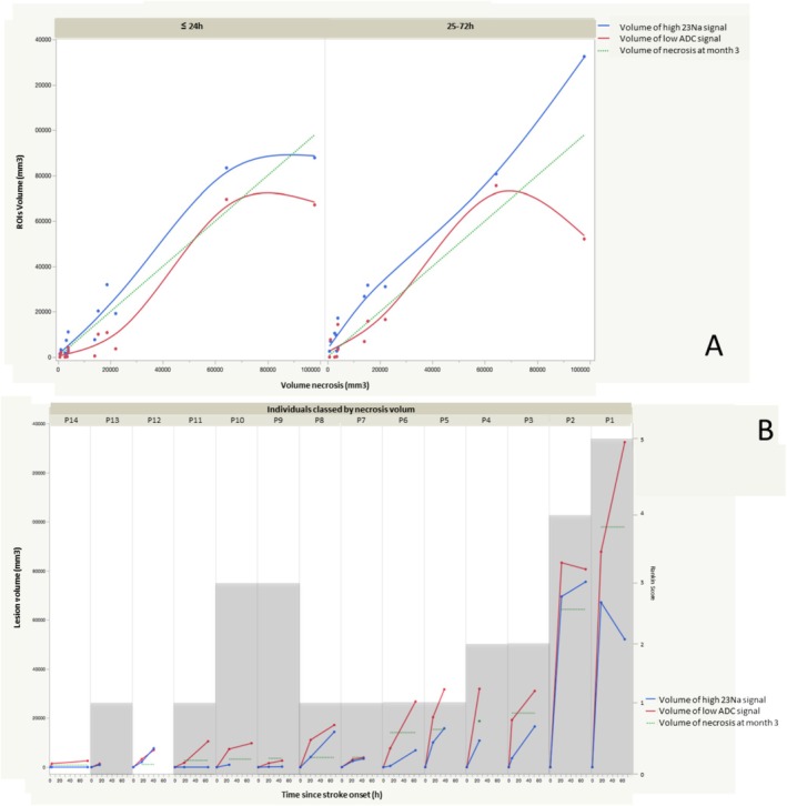
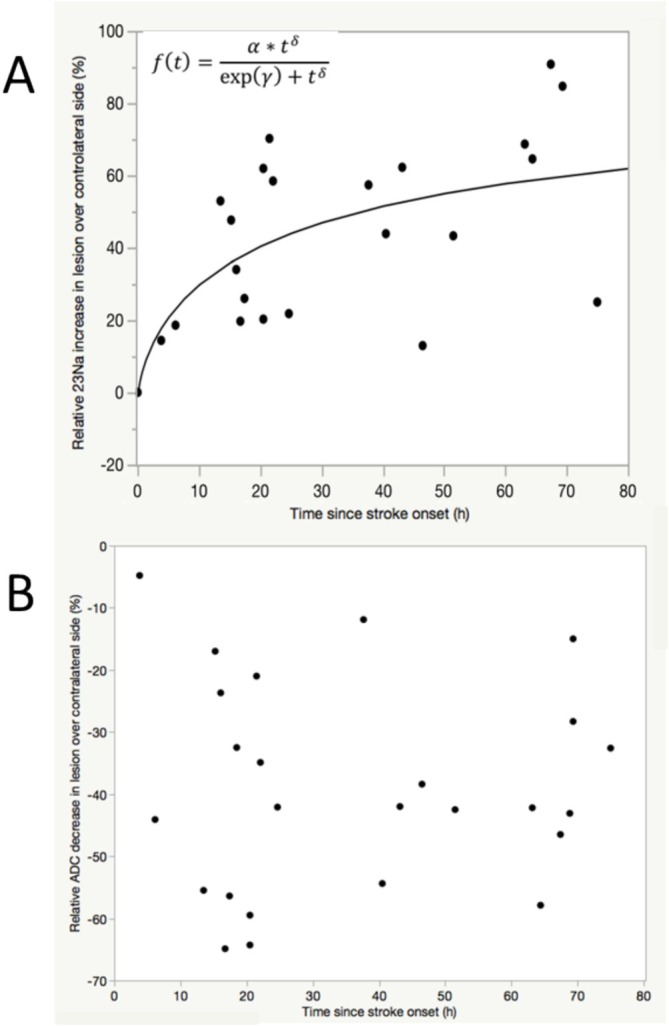
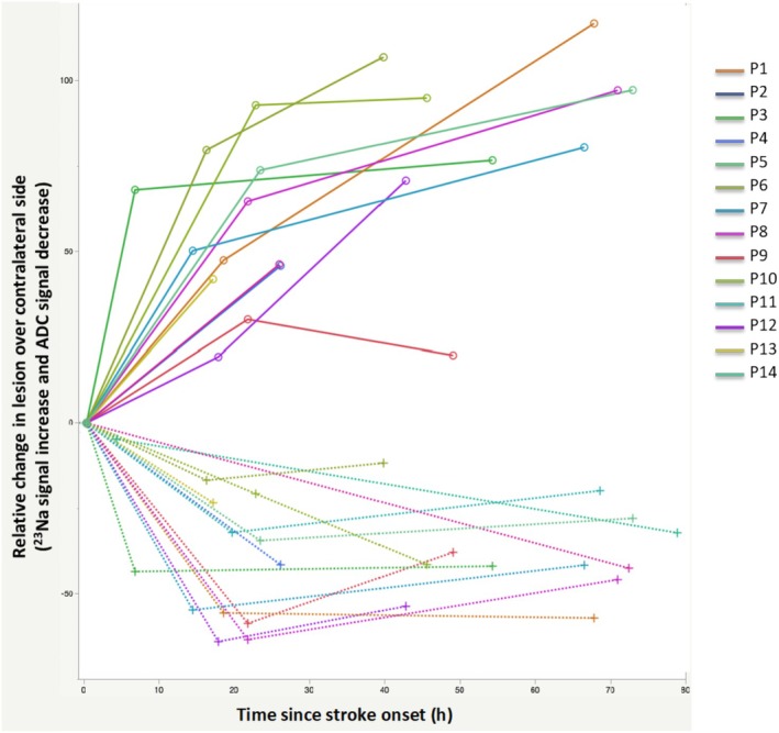
Cerebral imaging is crucial in the diagnosis and treatment algorithm of acute stroke to determine salvageable brain tissue. While diffusion MRI is commonly used to define the ischemic core, it cannot reliably distinguish irreversibly damaged from salvageable tissue. We investigated the added value of 23Na MRI to define irreversible necrotic tissue after a stroke. Fifteen patients with acute stroke involving medial cerebral artery occlusion were longitudinally explored with conventional and 23Na MRI within 24 h, 70 h following stroke and at 3 months to characterize the necrotic area. Time-courses of sodium accumulations were observed within regions presenting with or spared by cytotoxic/ionic edema and converting or not to necrosis. Dynamics of sodium accumulations were very different across subjects. At the group level, time-courses of sodium signal in cytotoxic edema showed a non-linear increase with an upper asymptote of 59 ± 6%% relative to the contralateral hemisphere. Regions with a larger early increase in 23Na signal (ionic edema) showed a non-linear accumulation during the first 70 h and were associated with subsequent necrosis at month 3. Some of the regions with no ionic edema during the first 70 h became necrotic at month 3, showing that pejorative pathophysiological processes could worsen after 70 h following attack. Final necrotic volume was well predicted by the cytotoxic volume (ADC decrease) during the first 24 h, and by the volume of ionic edema during the subacute period (25-70 h) following attack. The regions showing ionic edema showed a non-linear increase of 23Na signal during the first 70 h, with larger sodium accumulations in regions converting to necrosis at month 3. It may be of interest to consider the role of ionic edema imaging in the 70 h after stroke and reperfusion, with a view to better understand stroke pathophysiology. Sodium MRI could add complementary information about the fate of cell necrosis within low ADC signal regions.
期刊介绍:
NMR in Biomedicine is a journal devoted to the publication of original full-length papers, rapid communications and review articles describing the development of magnetic resonance spectroscopy or imaging methods or their use to investigate physiological, biochemical, biophysical or medical problems. Topics for submitted papers should be in one of the following general categories: (a) development of methods and instrumentation for MR of biological systems; (b) studies of normal or diseased organs, tissues or cells; (c) diagnosis or treatment of disease. Reports may cover work on patients or healthy human subjects, in vivo animal experiments, studies of isolated organs or cultured cells, analysis of tissue extracts, NMR theory, experimental techniques, or instrumentation.

 求助内容:
求助内容: 应助结果提醒方式:


