{"title":"优化晶状体切除术中人工晶状体度数计算的眼轴长度估算。","authors":"Sukhum Silpa-Archa, Chitchanok Samanwongthai, Variya Nganthavee, Korawin Charoensuk","doi":"10.1186/s40942-025-00666-5","DOIUrl":null,"url":null,"abstract":"<p><strong>Background: </strong>To evaluate methods of preoperative axial length (AL) estimation for intraocular lens (IOL) power calculation in patients with macula-off rhegmatogenous retinal detachment (RRD). These methods included optical biometry, A-scan biometry, and novel decision algorithms.</p><p><strong>Methods: </strong>A retrospective analysis of prospectively collected data was conducted at a tertiary hospital from January 2018 to December 2023. Preoperative and postoperative AL measurements were obtained using optical biometry (IOL Master 700, Zeiss, Germany) and A-scan biometry (VuMAX, Sonomed, USA). The primary outcome was the mean absolute prediction error (MAE) between postoperative AL and preoperative estimates generated by five methods, including two novel algorithms.</p><p><strong>Results: </strong>The study included 56 patients (56 eyes). The lowest MAE was achieved using the simple algorithm (0.31 ± 0.55 mm), followed by the AL of the fellow eye measured via IOL Master (0.34 ± 0.60 mm), and the advanced algorithm (0.36 ± 0.62 mm). A Kruskal-Wallis H test found no statistically significant difference in MAE across the five methods (P = 0.118). Bland-Altman analysis demonstrated good agreement between preoperative and postoperative AL measurements obtained with the IOL Master.</p><p><strong>Conclusion: </strong>For patients undergoing phacovitrectomy for macula-off RRD, the simple algorithm provides accurate AL estimation for IOL power calculation. In cases where AL measurement of the affected eye is not feasible using the IOL Master, the fellow eye's AL is a reliable alternative.</p>","PeriodicalId":14289,"journal":{"name":"International Journal of Retina and Vitreous","volume":"11 1","pages":"39"},"PeriodicalIF":2.4000,"publicationDate":"2025-04-02","publicationTypes":"Journal Article","fieldsOfStudy":null,"isOpenAccess":false,"openAccessPdf":"https://www.ncbi.nlm.nih.gov/pmc/articles/PMC11967035/pdf/","citationCount":"0","resultStr":"{\"title\":\"Optimizing axial length estimation for intraocular lens power calculation in phacovitrectomy for macula-off retinal detachment.\",\"authors\":\"Sukhum Silpa-Archa, Chitchanok Samanwongthai, Variya Nganthavee, Korawin Charoensuk\",\"doi\":\"10.1186/s40942-025-00666-5\",\"DOIUrl\":null,\"url\":null,\"abstract\":\"<p><strong>Background: </strong>To evaluate methods of preoperative axial length (AL) estimation for intraocular lens (IOL) power calculation in patients with macula-off rhegmatogenous retinal detachment (RRD). These methods included optical biometry, A-scan biometry, and novel decision algorithms.</p><p><strong>Methods: </strong>A retrospective analysis of prospectively collected data was conducted at a tertiary hospital from January 2018 to December 2023. Preoperative and postoperative AL measurements were obtained using optical biometry (IOL Master 700, Zeiss, Germany) and A-scan biometry (VuMAX, Sonomed, USA). The primary outcome was the mean absolute prediction error (MAE) between postoperative AL and preoperative estimates generated by five methods, including two novel algorithms.</p><p><strong>Results: </strong>The study included 56 patients (56 eyes). The lowest MAE was achieved using the simple algorithm (0.31 ± 0.55 mm), followed by the AL of the fellow eye measured via IOL Master (0.34 ± 0.60 mm), and the advanced algorithm (0.36 ± 0.62 mm). A Kruskal-Wallis H test found no statistically significant difference in MAE across the five methods (P = 0.118). Bland-Altman analysis demonstrated good agreement between preoperative and postoperative AL measurements obtained with the IOL Master.</p><p><strong>Conclusion: </strong>For patients undergoing phacovitrectomy for macula-off RRD, the simple algorithm provides accurate AL estimation for IOL power calculation. In cases where AL measurement of the affected eye is not feasible using the IOL Master, the fellow eye's AL is a reliable alternative.</p>\",\"PeriodicalId\":14289,\"journal\":{\"name\":\"International Journal of Retina and Vitreous\",\"volume\":\"11 1\",\"pages\":\"39\"},\"PeriodicalIF\":2.4000,\"publicationDate\":\"2025-04-02\",\"publicationTypes\":\"Journal Article\",\"fieldsOfStudy\":null,\"isOpenAccess\":false,\"openAccessPdf\":\"https://www.ncbi.nlm.nih.gov/pmc/articles/PMC11967035/pdf/\",\"citationCount\":\"0\",\"resultStr\":null,\"platform\":\"Semanticscholar\",\"paperid\":null,\"PeriodicalName\":\"International Journal of Retina and Vitreous\",\"FirstCategoryId\":\"1085\",\"ListUrlMain\":\"https://doi.org/10.1186/s40942-025-00666-5\",\"RegionNum\":0,\"RegionCategory\":null,\"ArticlePicture\":[],\"TitleCN\":null,\"AbstractTextCN\":null,\"PMCID\":null,\"EPubDate\":\"\",\"PubModel\":\"\",\"JCR\":\"Q2\",\"JCRName\":\"OPHTHALMOLOGY\",\"Score\":null,\"Total\":0}","platform":"Semanticscholar","paperid":null,"PeriodicalName":"International Journal of Retina and Vitreous","FirstCategoryId":"1085","ListUrlMain":"https://doi.org/10.1186/s40942-025-00666-5","RegionNum":0,"RegionCategory":null,"ArticlePicture":[],"TitleCN":null,"AbstractTextCN":null,"PMCID":null,"EPubDate":"","PubModel":"","JCR":"Q2","JCRName":"OPHTHALMOLOGY","Score":null,"Total":0}
Optimizing axial length estimation for intraocular lens power calculation in phacovitrectomy for macula-off retinal detachment.
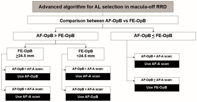
Background: To evaluate methods of preoperative axial length (AL) estimation for intraocular lens (IOL) power calculation in patients with macula-off rhegmatogenous retinal detachment (RRD). These methods included optical biometry, A-scan biometry, and novel decision algorithms.
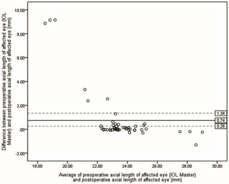
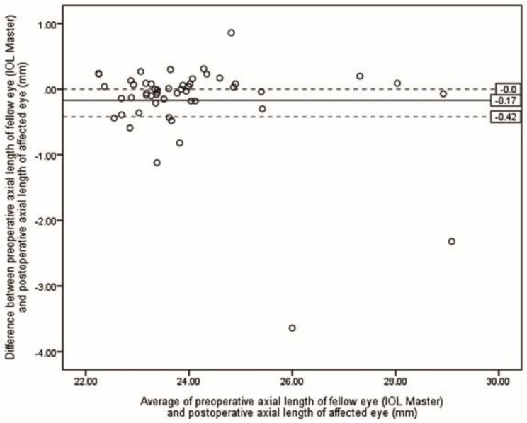
Methods: A retrospective analysis of prospectively collected data was conducted at a tertiary hospital from January 2018 to December 2023. Preoperative and postoperative AL measurements were obtained using optical biometry (IOL Master 700, Zeiss, Germany) and A-scan biometry (VuMAX, Sonomed, USA). The primary outcome was the mean absolute prediction error (MAE) between postoperative AL and preoperative estimates generated by five methods, including two novel algorithms.
Results: The study included 56 patients (56 eyes). The lowest MAE was achieved using the simple algorithm (0.31 ± 0.55 mm), followed by the AL of the fellow eye measured via IOL Master (0.34 ± 0.60 mm), and the advanced algorithm (0.36 ± 0.62 mm). A Kruskal-Wallis H test found no statistically significant difference in MAE across the five methods (P = 0.118). Bland-Altman analysis demonstrated good agreement between preoperative and postoperative AL measurements obtained with the IOL Master.
Conclusion: For patients undergoing phacovitrectomy for macula-off RRD, the simple algorithm provides accurate AL estimation for IOL power calculation. In cases where AL measurement of the affected eye is not feasible using the IOL Master, the fellow eye's AL is a reliable alternative.
期刊介绍:
International Journal of Retina and Vitreous focuses on the ophthalmic subspecialty of vitreoretinal disorders. The journal presents original articles on new approaches to diagnosis, outcomes of clinical trials, innovations in pharmacological therapy and surgical techniques, as well as basic science advances that impact clinical practice. Topical areas include, but are not limited to: -Imaging of the retina, choroid and vitreous -Innovations in optical coherence tomography (OCT) -Small-gauge vitrectomy, retinal detachment, chromovitrectomy -Electroretinography (ERG), microperimetry, other functional tests -Intraocular tumors -Retinal pharmacotherapy & drug delivery -Diabetic retinopathy & other vascular diseases -Age-related macular degeneration (AMD) & other macular entities

 求助内容:
求助内容: 应助结果提醒方式:


