Gabriela O'Toole Bom Braga, Robert Zboray, Annapaola Parrilli, Franca Wagner
{"title":"体绘制技术和高分辨率微ct:耳蜗解剖的三维探索。","authors":"Gabriela O'Toole Bom Braga, Robert Zboray, Annapaola Parrilli, Franca Wagner","doi":"10.1007/s00405-025-09360-6","DOIUrl":null,"url":null,"abstract":"<p><strong>Purpose: </strong>Given its unique anatomical position and the amalgamation of bony and soft tissues within the cochlea, exploring its intricacies poses persistent challenges. Histopathology remains the gold standard in research, but given its inherent limitations, there is a clear need for innovative alternatives. The integration of microCT technology with advanced volume rendering techniques emerges as a promising approach for overcoming the hurdles associated with anatomical investigations of the cochlea.</p><p><strong>Methods: </strong>We seamlessly integrated high-resolution microCT cochlear images with medical imaging analysis software to create detailed 3D anatomical images of the human cochlea without the need of sample processing.</p><p><strong>Results: </strong>Volume rendering allowed a multiplanar, non-destructive, detailed anatomical evaluation of the human cochlea, including its capillary system, as well as soft tissue visualization at single-micron resolution in 3D.</p><p><strong>Conclusion: </strong>The use of volume rendering in cochlear anatomical studies is underexplored despite the prevalence of 3D reconstruction. This technique presents a promising avenue for scientific investigation, providing researchers with unprecedented insights that can potentially benefit patients with hearing disorders.</p>","PeriodicalId":11952,"journal":{"name":"European Archives of Oto-Rhino-Laryngology","volume":" ","pages":"4497-4504"},"PeriodicalIF":2.2000,"publicationDate":"2025-09-01","publicationTypes":"Journal Article","fieldsOfStudy":null,"isOpenAccess":false,"openAccessPdf":"https://www.ncbi.nlm.nih.gov/pmc/articles/PMC12423228/pdf/","citationCount":"0","resultStr":"{\"title\":\"Volume rendering technique and high-resolution microCT: 3D exploration of the cochlear anatomy.\",\"authors\":\"Gabriela O'Toole Bom Braga, Robert Zboray, Annapaola Parrilli, Franca Wagner\",\"doi\":\"10.1007/s00405-025-09360-6\",\"DOIUrl\":null,\"url\":null,\"abstract\":\"<p><strong>Purpose: </strong>Given its unique anatomical position and the amalgamation of bony and soft tissues within the cochlea, exploring its intricacies poses persistent challenges. Histopathology remains the gold standard in research, but given its inherent limitations, there is a clear need for innovative alternatives. The integration of microCT technology with advanced volume rendering techniques emerges as a promising approach for overcoming the hurdles associated with anatomical investigations of the cochlea.</p><p><strong>Methods: </strong>We seamlessly integrated high-resolution microCT cochlear images with medical imaging analysis software to create detailed 3D anatomical images of the human cochlea without the need of sample processing.</p><p><strong>Results: </strong>Volume rendering allowed a multiplanar, non-destructive, detailed anatomical evaluation of the human cochlea, including its capillary system, as well as soft tissue visualization at single-micron resolution in 3D.</p><p><strong>Conclusion: </strong>The use of volume rendering in cochlear anatomical studies is underexplored despite the prevalence of 3D reconstruction. This technique presents a promising avenue for scientific investigation, providing researchers with unprecedented insights that can potentially benefit patients with hearing disorders.</p>\",\"PeriodicalId\":11952,\"journal\":{\"name\":\"European Archives of Oto-Rhino-Laryngology\",\"volume\":\" \",\"pages\":\"4497-4504\"},\"PeriodicalIF\":2.2000,\"publicationDate\":\"2025-09-01\",\"publicationTypes\":\"Journal Article\",\"fieldsOfStudy\":null,\"isOpenAccess\":false,\"openAccessPdf\":\"https://www.ncbi.nlm.nih.gov/pmc/articles/PMC12423228/pdf/\",\"citationCount\":\"0\",\"resultStr\":null,\"platform\":\"Semanticscholar\",\"paperid\":null,\"PeriodicalName\":\"European Archives of Oto-Rhino-Laryngology\",\"FirstCategoryId\":\"3\",\"ListUrlMain\":\"https://doi.org/10.1007/s00405-025-09360-6\",\"RegionNum\":3,\"RegionCategory\":\"医学\",\"ArticlePicture\":[],\"TitleCN\":null,\"AbstractTextCN\":null,\"PMCID\":null,\"EPubDate\":\"2025/4/3 0:00:00\",\"PubModel\":\"Epub\",\"JCR\":\"Q2\",\"JCRName\":\"OTORHINOLARYNGOLOGY\",\"Score\":null,\"Total\":0}","platform":"Semanticscholar","paperid":null,"PeriodicalName":"European Archives of Oto-Rhino-Laryngology","FirstCategoryId":"3","ListUrlMain":"https://doi.org/10.1007/s00405-025-09360-6","RegionNum":3,"RegionCategory":"医学","ArticlePicture":[],"TitleCN":null,"AbstractTextCN":null,"PMCID":null,"EPubDate":"2025/4/3 0:00:00","PubModel":"Epub","JCR":"Q2","JCRName":"OTORHINOLARYNGOLOGY","Score":null,"Total":0}
Volume rendering technique and high-resolution microCT: 3D exploration of the cochlear anatomy.
Purpose: Given its unique anatomical position and the amalgamation of bony and soft tissues within the cochlea, exploring its intricacies poses persistent challenges. Histopathology remains the gold standard in research, but given its inherent limitations, there is a clear need for innovative alternatives. The integration of microCT technology with advanced volume rendering techniques emerges as a promising approach for overcoming the hurdles associated with anatomical investigations of the cochlea.
Methods: We seamlessly integrated high-resolution microCT cochlear images with medical imaging analysis software to create detailed 3D anatomical images of the human cochlea without the need of sample processing.
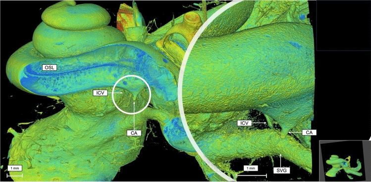
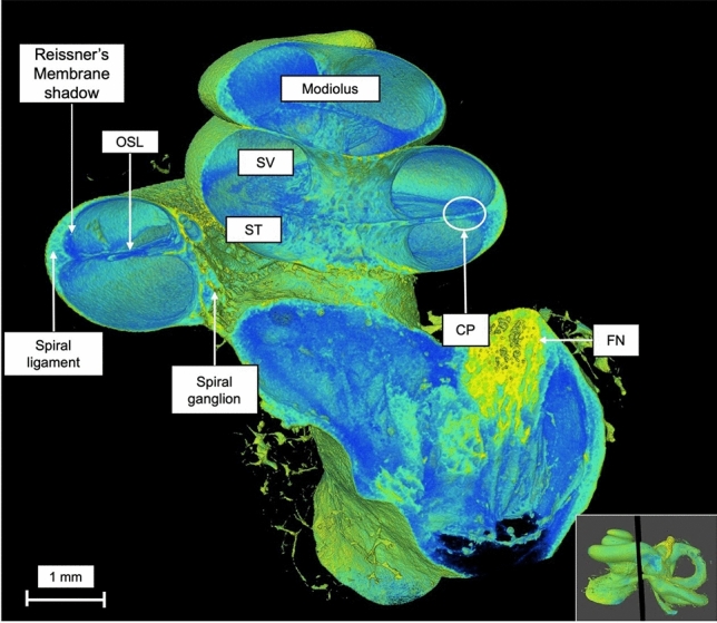
Results: Volume rendering allowed a multiplanar, non-destructive, detailed anatomical evaluation of the human cochlea, including its capillary system, as well as soft tissue visualization at single-micron resolution in 3D.
Conclusion: The use of volume rendering in cochlear anatomical studies is underexplored despite the prevalence of 3D reconstruction. This technique presents a promising avenue for scientific investigation, providing researchers with unprecedented insights that can potentially benefit patients with hearing disorders.
期刊介绍:
Official Journal of
European Union of Medical Specialists – ORL Section and Board
Official Journal of Confederation of European Oto-Rhino-Laryngology Head and Neck Surgery
"European Archives of Oto-Rhino-Laryngology" publishes original clinical reports and clinically relevant experimental studies, as well as short communications presenting new results of special interest. With peer review by a respected international editorial board and prompt English-language publication, the journal provides rapid dissemination of information by authors from around the world. This particular feature makes it the journal of choice for readers who want to be informed about the continuing state of the art concerning basic sciences and the diagnosis and management of diseases of the head and neck on an international level.
European Archives of Oto-Rhino-Laryngology was founded in 1864 as "Archiv für Ohrenheilkunde" by A. von Tröltsch, A. Politzer and H. Schwartze.

 求助内容:
求助内容: 应助结果提醒方式:


