Jun Liu, Huanhua Wu, Dabin Ren, Hao Huang, Xinyue Chen, Liqiu Liu, Yongtao Wang, Guoyu Wang
{"title":"动脉期CT放射组学无创预测胰腺实体性假乳头状肿瘤Ki-67增殖指数。","authors":"Jun Liu, Huanhua Wu, Dabin Ren, Hao Huang, Xinyue Chen, Liqiu Liu, Yongtao Wang, Guoyu Wang","doi":"10.1007/s00261-025-04921-z","DOIUrl":null,"url":null,"abstract":"<div><h3>Background</h3><p>This study aimed to preoperatively predict Ki-67 proliferation levels in patients with pancreatic solid pseudopapillary neoplasm (pSPN) using radiomics features extracted from arterial phase helical CT images.</p><h3>Methods</h3><p>We retrospectively analyzed 92 patients (Ningbo Medical Center Lihuili Hospital: <i>n</i> = 64, Taizhou Central Hospital: <i>n</i> = 28) with pathologically confirmed pSPN from June 2015 to June 2023. Ki-67 positivity > 3% was considered high. Radiomics features were extracted using PyRadiomics, with patients from training cohort (<i>n</i> = 64) and validation cohort (<i>n</i> = 28). A radiomics signature was constructed, and a CT radiomics score (CTscore) was calculated. Deep learning models were employed for prediction, with early stopping to prevent overfitting.</p><h3>Results</h3><p>Seven key radiomics features were selected via LASSO regression with cross-validation. The deep learning model demonstrated improved accuracy with demographics and CTscore, with key features such as Morphology and CTscore contributing significantly to predictive accuracy. The best-performing models, including GBM and deep learning algorithms, achieved high predictive performance with an AUC of up to 0.946 in the training cohort.</p><h3>Conclusions</h3><p>We developed a robust deep learning-based radiomics model using arterial phase CT images to predict Ki-67 levels in pSPN patients, identifying CTscore and Morphology as key predictors. This non-invasive approach has potential utility in guiding personalized preoperative treatment strategies.</p><h3>Clinical trial number</h3><p>Not applicable.</p></div>","PeriodicalId":7126,"journal":{"name":"Abdominal Radiology","volume":"50 10","pages":"4635 - 4645"},"PeriodicalIF":2.2000,"publicationDate":"2025-04-03","publicationTypes":"Journal Article","fieldsOfStudy":null,"isOpenAccess":false,"openAccessPdf":"","citationCount":"0","resultStr":"{\"title\":\"Arterial phase CT radiomics for non-invasive prediction of Ki-67 proliferation index in pancreatic solid pseudopapillary neoplasms\",\"authors\":\"Jun Liu, Huanhua Wu, Dabin Ren, Hao Huang, Xinyue Chen, Liqiu Liu, Yongtao Wang, Guoyu Wang\",\"doi\":\"10.1007/s00261-025-04921-z\",\"DOIUrl\":null,\"url\":null,\"abstract\":\"<div><h3>Background</h3><p>This study aimed to preoperatively predict Ki-67 proliferation levels in patients with pancreatic solid pseudopapillary neoplasm (pSPN) using radiomics features extracted from arterial phase helical CT images.</p><h3>Methods</h3><p>We retrospectively analyzed 92 patients (Ningbo Medical Center Lihuili Hospital: <i>n</i> = 64, Taizhou Central Hospital: <i>n</i> = 28) with pathologically confirmed pSPN from June 2015 to June 2023. Ki-67 positivity > 3% was considered high. Radiomics features were extracted using PyRadiomics, with patients from training cohort (<i>n</i> = 64) and validation cohort (<i>n</i> = 28). A radiomics signature was constructed, and a CT radiomics score (CTscore) was calculated. Deep learning models were employed for prediction, with early stopping to prevent overfitting.</p><h3>Results</h3><p>Seven key radiomics features were selected via LASSO regression with cross-validation. The deep learning model demonstrated improved accuracy with demographics and CTscore, with key features such as Morphology and CTscore contributing significantly to predictive accuracy. The best-performing models, including GBM and deep learning algorithms, achieved high predictive performance with an AUC of up to 0.946 in the training cohort.</p><h3>Conclusions</h3><p>We developed a robust deep learning-based radiomics model using arterial phase CT images to predict Ki-67 levels in pSPN patients, identifying CTscore and Morphology as key predictors. This non-invasive approach has potential utility in guiding personalized preoperative treatment strategies.</p><h3>Clinical trial number</h3><p>Not applicable.</p></div>\",\"PeriodicalId\":7126,\"journal\":{\"name\":\"Abdominal Radiology\",\"volume\":\"50 10\",\"pages\":\"4635 - 4645\"},\"PeriodicalIF\":2.2000,\"publicationDate\":\"2025-04-03\",\"publicationTypes\":\"Journal Article\",\"fieldsOfStudy\":null,\"isOpenAccess\":false,\"openAccessPdf\":\"\",\"citationCount\":\"0\",\"resultStr\":null,\"platform\":\"Semanticscholar\",\"paperid\":null,\"PeriodicalName\":\"Abdominal Radiology\",\"FirstCategoryId\":\"3\",\"ListUrlMain\":\"https://link.springer.com/article/10.1007/s00261-025-04921-z\",\"RegionNum\":3,\"RegionCategory\":\"医学\",\"ArticlePicture\":[],\"TitleCN\":null,\"AbstractTextCN\":null,\"PMCID\":null,\"EPubDate\":\"\",\"PubModel\":\"\",\"JCR\":\"Q2\",\"JCRName\":\"RADIOLOGY, NUCLEAR MEDICINE & MEDICAL IMAGING\",\"Score\":null,\"Total\":0}","platform":"Semanticscholar","paperid":null,"PeriodicalName":"Abdominal Radiology","FirstCategoryId":"3","ListUrlMain":"https://link.springer.com/article/10.1007/s00261-025-04921-z","RegionNum":3,"RegionCategory":"医学","ArticlePicture":[],"TitleCN":null,"AbstractTextCN":null,"PMCID":null,"EPubDate":"","PubModel":"","JCR":"Q2","JCRName":"RADIOLOGY, NUCLEAR MEDICINE & MEDICAL IMAGING","Score":null,"Total":0}
Arterial phase CT radiomics for non-invasive prediction of Ki-67 proliferation index in pancreatic solid pseudopapillary neoplasms
Background
This study aimed to preoperatively predict Ki-67 proliferation levels in patients with pancreatic solid pseudopapillary neoplasm (pSPN) using radiomics features extracted from arterial phase helical CT images.
Methods
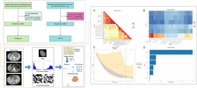
We retrospectively analyzed 92 patients (Ningbo Medical Center Lihuili Hospital: n = 64, Taizhou Central Hospital: n = 28) with pathologically confirmed pSPN from June 2015 to June 2023. Ki-67 positivity > 3% was considered high. Radiomics features were extracted using PyRadiomics, with patients from training cohort (n = 64) and validation cohort (n = 28). A radiomics signature was constructed, and a CT radiomics score (CTscore) was calculated. Deep learning models were employed for prediction, with early stopping to prevent overfitting.
Results
Seven key radiomics features were selected via LASSO regression with cross-validation. The deep learning model demonstrated improved accuracy with demographics and CTscore, with key features such as Morphology and CTscore contributing significantly to predictive accuracy. The best-performing models, including GBM and deep learning algorithms, achieved high predictive performance with an AUC of up to 0.946 in the training cohort.
Conclusions
We developed a robust deep learning-based radiomics model using arterial phase CT images to predict Ki-67 levels in pSPN patients, identifying CTscore and Morphology as key predictors. This non-invasive approach has potential utility in guiding personalized preoperative treatment strategies.
期刊介绍:
Abdominal Radiology seeks to meet the professional needs of the abdominal radiologist by publishing clinically pertinent original, review and practice related articles on the gastrointestinal and genitourinary tracts and abdominal interventional and radiologic procedures. Case reports are generally not accepted unless they are the first report of a new disease or condition, or part of a special solicited section.
Reasons to Publish Your Article in Abdominal Radiology:
· Official journal of the Society of Abdominal Radiology (SAR)
· Published in Cooperation with:
European Society of Gastrointestinal and Abdominal Radiology (ESGAR)
European Society of Urogenital Radiology (ESUR)
Asian Society of Abdominal Radiology (ASAR)
· Efficient handling and Expeditious review
· Author feedback is provided in a mentoring style
· Global readership
· Readers can earn CME credits


 求助内容:
求助内容: 应助结果提醒方式:


