İlhan Özdemir, Ayfer Şanli Aktaş, Mehmet Cudi Tuncer
{"title":"百里醌和阿霉素对OVCAR3人卵巢腺癌细胞EGFR/FOXP3信号通路影响的研究","authors":"İlhan Özdemir, Ayfer Şanli Aktaş, Mehmet Cudi Tuncer","doi":"10.1590/acb401725","DOIUrl":null,"url":null,"abstract":"<p><strong>Purpose: </strong>To investigate the cytotoxic and apoptotic effects of the combination of doxorubicin (Dox) and thymoquinone (TQ) on ovarian adenocarcinoma cells (OVCAR3) via the EGFR/FOXP3 signaling pathway.</p><p><strong>Methods: </strong>We used human OVCAR3 and human skin keratinocyte cells (HaCaT). Different concentrations of TQ and Dox were applied to the cells for 24, 48, and 72 hours, and the cytotoxicity level was determined via the MTT method. Expression levels of EGFR/FOXP3 for cell proliferation and apoptosis were determined by quantitative reverse transcription polymerase chain reaction (RT-qPCR) and Western blot analysis. The colony counting was performed after DAPI staining, and the effect on cell proliferation was determined.</p><p><strong>Results: </strong>Cytotoxicity was found to be highest with TQ and Dox treatments, and cell migration was prevented, especially in the group that received combined TQ and Dox treatment. Moreover, using RT-qPCR analysis, activity in the EGFR and FOXP3 pathway was found to be downregulated the most with TQ, and the amount of protein decreased with TQ and Dox.</p><p><strong>Conclusions: </strong>The findings showed that the greatest cytotoxic effect and the most apoptosis occurred during TQ treatment. Additionally, it was determined that a significant decrease in EGFR and FOXP3 levels occurred with the application of TQ and Dox.</p>","PeriodicalId":93850,"journal":{"name":"Acta cirurgica brasileira","volume":"40 ","pages":"e401725"},"PeriodicalIF":1.3000,"publicationDate":"2025-03-31","publicationTypes":"Journal Article","fieldsOfStudy":null,"isOpenAccess":false,"openAccessPdf":"https://www.ncbi.nlm.nih.gov/pmc/articles/PMC11960576/pdf/","citationCount":"0","resultStr":"{\"title\":\"Investigation of the effect of thymoquinone and doxorubicin on the EGFR/FOXP3 signaling pathway in OVCAR3 human ovarian adenocarcinoma cells.\",\"authors\":\"İlhan Özdemir, Ayfer Şanli Aktaş, Mehmet Cudi Tuncer\",\"doi\":\"10.1590/acb401725\",\"DOIUrl\":null,\"url\":null,\"abstract\":\"<p><strong>Purpose: </strong>To investigate the cytotoxic and apoptotic effects of the combination of doxorubicin (Dox) and thymoquinone (TQ) on ovarian adenocarcinoma cells (OVCAR3) via the EGFR/FOXP3 signaling pathway.</p><p><strong>Methods: </strong>We used human OVCAR3 and human skin keratinocyte cells (HaCaT). Different concentrations of TQ and Dox were applied to the cells for 24, 48, and 72 hours, and the cytotoxicity level was determined via the MTT method. Expression levels of EGFR/FOXP3 for cell proliferation and apoptosis were determined by quantitative reverse transcription polymerase chain reaction (RT-qPCR) and Western blot analysis. The colony counting was performed after DAPI staining, and the effect on cell proliferation was determined.</p><p><strong>Results: </strong>Cytotoxicity was found to be highest with TQ and Dox treatments, and cell migration was prevented, especially in the group that received combined TQ and Dox treatment. Moreover, using RT-qPCR analysis, activity in the EGFR and FOXP3 pathway was found to be downregulated the most with TQ, and the amount of protein decreased with TQ and Dox.</p><p><strong>Conclusions: </strong>The findings showed that the greatest cytotoxic effect and the most apoptosis occurred during TQ treatment. Additionally, it was determined that a significant decrease in EGFR and FOXP3 levels occurred with the application of TQ and Dox.</p>\",\"PeriodicalId\":93850,\"journal\":{\"name\":\"Acta cirurgica brasileira\",\"volume\":\"40 \",\"pages\":\"e401725\"},\"PeriodicalIF\":1.3000,\"publicationDate\":\"2025-03-31\",\"publicationTypes\":\"Journal Article\",\"fieldsOfStudy\":null,\"isOpenAccess\":false,\"openAccessPdf\":\"https://www.ncbi.nlm.nih.gov/pmc/articles/PMC11960576/pdf/\",\"citationCount\":\"0\",\"resultStr\":null,\"platform\":\"Semanticscholar\",\"paperid\":null,\"PeriodicalName\":\"Acta cirurgica brasileira\",\"FirstCategoryId\":\"1085\",\"ListUrlMain\":\"https://doi.org/10.1590/acb401725\",\"RegionNum\":0,\"RegionCategory\":null,\"ArticlePicture\":[],\"TitleCN\":null,\"AbstractTextCN\":null,\"PMCID\":null,\"EPubDate\":\"2025/1/1 0:00:00\",\"PubModel\":\"eCollection\",\"JCR\":\"\",\"JCRName\":\"\",\"Score\":null,\"Total\":0}","platform":"Semanticscholar","paperid":null,"PeriodicalName":"Acta cirurgica brasileira","FirstCategoryId":"1085","ListUrlMain":"https://doi.org/10.1590/acb401725","RegionNum":0,"RegionCategory":null,"ArticlePicture":[],"TitleCN":null,"AbstractTextCN":null,"PMCID":null,"EPubDate":"2025/1/1 0:00:00","PubModel":"eCollection","JCR":"","JCRName":"","Score":null,"Total":0}
Investigation of the effect of thymoquinone and doxorubicin on the EGFR/FOXP3 signaling pathway in OVCAR3 human ovarian adenocarcinoma cells.
Purpose: To investigate the cytotoxic and apoptotic effects of the combination of doxorubicin (Dox) and thymoquinone (TQ) on ovarian adenocarcinoma cells (OVCAR3) via the EGFR/FOXP3 signaling pathway.
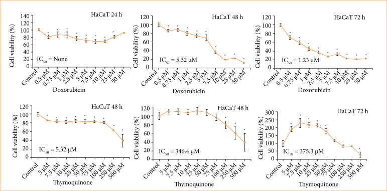
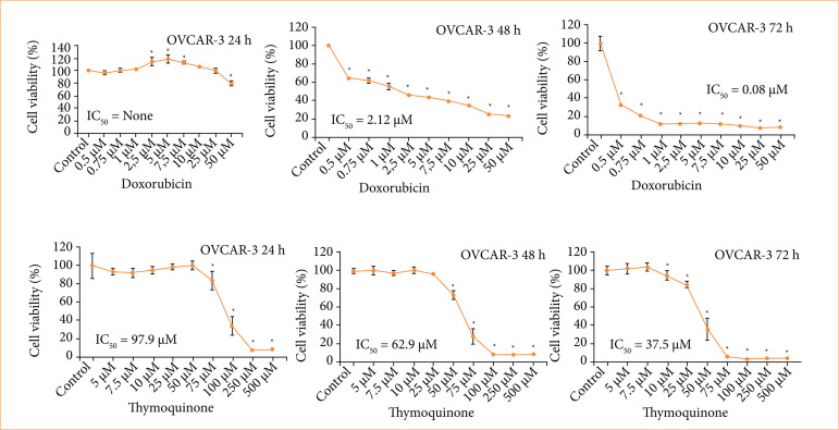
Methods: We used human OVCAR3 and human skin keratinocyte cells (HaCaT). Different concentrations of TQ and Dox were applied to the cells for 24, 48, and 72 hours, and the cytotoxicity level was determined via the MTT method. Expression levels of EGFR/FOXP3 for cell proliferation and apoptosis were determined by quantitative reverse transcription polymerase chain reaction (RT-qPCR) and Western blot analysis. The colony counting was performed after DAPI staining, and the effect on cell proliferation was determined.
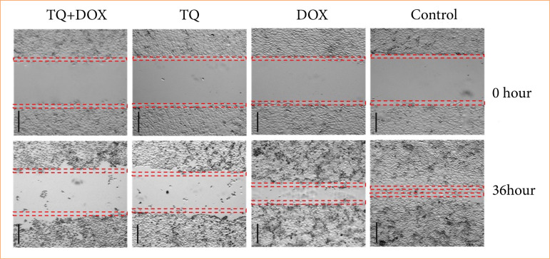
Results: Cytotoxicity was found to be highest with TQ and Dox treatments, and cell migration was prevented, especially in the group that received combined TQ and Dox treatment. Moreover, using RT-qPCR analysis, activity in the EGFR and FOXP3 pathway was found to be downregulated the most with TQ, and the amount of protein decreased with TQ and Dox.
Conclusions: The findings showed that the greatest cytotoxic effect and the most apoptosis occurred during TQ treatment. Additionally, it was determined that a significant decrease in EGFR and FOXP3 levels occurred with the application of TQ and Dox.

 求助内容:
求助内容: 应助结果提醒方式:


