{"title":"间歇性缺氧刺激可促进缺氧诱导因子-1α的有效表达,并在动物骨关节炎模型中发挥软骨保护作用。","authors":"Ryota Cha, Shuji Nakagawa, Yuji Arai, Atsuo Inoue, Naoki Okubo, Yuta Fujii, Kenta Kaihara, Kei Nakamura, Tsunao Kishida, Osam Mazda, Kenji Takahashi","doi":"10.1371/journal.pone.0319976","DOIUrl":null,"url":null,"abstract":"<p><p>Hypoxia-inducible factor-1α plays an important role in the homeostasis of articular cartilage in hypoxic environments. Therefore, modulation of hypoxia-inducible factor-1α by regulating the oxygen environment could be a useful treatment for osteoarthritis. This study aimed to assess the chondroprotective effects of intermittent hypoxia on cultured chondrocytes and an animal model of osteoarthritis. In vitro, human chondrocytes were exposed to 2 h of hypoxic stimulation three times at 1-h intervals, and protein and gene expression of hypoxia-inducible factor-1α, ACAN, and cell viability was measured over time. In vivo, 8-week-old male Wistar rats were injected with monosodium iodoacetate to induce osteoarthritis and then reared in 12% hypoxia for 24 h, followed by 24 h in steady oxygen, repeated alternately for a total of 28 days. A histological analysis was performed on days 8 and 28. In the intermittent hypoxia group, each protein expression increased with each repeated hypoxic stimulation to human chondrocytes; finally, the protein level was significantly higher with intermittent hypoxia than with continuous hypoxic stimulation, cell viability was increased, and gene expression was not significantly increased. In the osteoarthritis animal model, for 8 days, there were stronger hypoxia-inducible factor-1α staining and no significant differences in articular cartilage destruction. Furthermore, for 28 days, there was significantly less articular cartilage destruction in the rat osteoarthritis model with intermittent hypoxia than with steady oxygen rearing. Intermittent hypoxia increased cartilage metabolism by increasing hypoxia-inducible factor-1α proteins in articular chondrocytes, which may be effective in preventing articular cartilage degeneration in a rat osteoarthritis model.</p>","PeriodicalId":20189,"journal":{"name":"PLoS ONE","volume":"20 4","pages":"e0319976"},"PeriodicalIF":2.6000,"publicationDate":"2025-04-01","publicationTypes":"Journal Article","fieldsOfStudy":null,"isOpenAccess":false,"openAccessPdf":"https://www.ncbi.nlm.nih.gov/pmc/articles/PMC11960973/pdf/","citationCount":"0","resultStr":"{\"title\":\"Intermittent hypoxic stimulation promotes efficient expression of Hypoxia-inducible factor-1α and exerts a chondroprotective effect in an animal osteoarthritis model.\",\"authors\":\"Ryota Cha, Shuji Nakagawa, Yuji Arai, Atsuo Inoue, Naoki Okubo, Yuta Fujii, Kenta Kaihara, Kei Nakamura, Tsunao Kishida, Osam Mazda, Kenji Takahashi\",\"doi\":\"10.1371/journal.pone.0319976\",\"DOIUrl\":null,\"url\":null,\"abstract\":\"<p><p>Hypoxia-inducible factor-1α plays an important role in the homeostasis of articular cartilage in hypoxic environments. Therefore, modulation of hypoxia-inducible factor-1α by regulating the oxygen environment could be a useful treatment for osteoarthritis. This study aimed to assess the chondroprotective effects of intermittent hypoxia on cultured chondrocytes and an animal model of osteoarthritis. In vitro, human chondrocytes were exposed to 2 h of hypoxic stimulation three times at 1-h intervals, and protein and gene expression of hypoxia-inducible factor-1α, ACAN, and cell viability was measured over time. In vivo, 8-week-old male Wistar rats were injected with monosodium iodoacetate to induce osteoarthritis and then reared in 12% hypoxia for 24 h, followed by 24 h in steady oxygen, repeated alternately for a total of 28 days. A histological analysis was performed on days 8 and 28. In the intermittent hypoxia group, each protein expression increased with each repeated hypoxic stimulation to human chondrocytes; finally, the protein level was significantly higher with intermittent hypoxia than with continuous hypoxic stimulation, cell viability was increased, and gene expression was not significantly increased. In the osteoarthritis animal model, for 8 days, there were stronger hypoxia-inducible factor-1α staining and no significant differences in articular cartilage destruction. Furthermore, for 28 days, there was significantly less articular cartilage destruction in the rat osteoarthritis model with intermittent hypoxia than with steady oxygen rearing. Intermittent hypoxia increased cartilage metabolism by increasing hypoxia-inducible factor-1α proteins in articular chondrocytes, which may be effective in preventing articular cartilage degeneration in a rat osteoarthritis model.</p>\",\"PeriodicalId\":20189,\"journal\":{\"name\":\"PLoS ONE\",\"volume\":\"20 4\",\"pages\":\"e0319976\"},\"PeriodicalIF\":2.6000,\"publicationDate\":\"2025-04-01\",\"publicationTypes\":\"Journal Article\",\"fieldsOfStudy\":null,\"isOpenAccess\":false,\"openAccessPdf\":\"https://www.ncbi.nlm.nih.gov/pmc/articles/PMC11960973/pdf/\",\"citationCount\":\"0\",\"resultStr\":null,\"platform\":\"Semanticscholar\",\"paperid\":null,\"PeriodicalName\":\"PLoS ONE\",\"FirstCategoryId\":\"103\",\"ListUrlMain\":\"https://doi.org/10.1371/journal.pone.0319976\",\"RegionNum\":3,\"RegionCategory\":\"综合性期刊\",\"ArticlePicture\":[],\"TitleCN\":null,\"AbstractTextCN\":null,\"PMCID\":null,\"EPubDate\":\"2025/1/1 0:00:00\",\"PubModel\":\"eCollection\",\"JCR\":\"Q1\",\"JCRName\":\"MULTIDISCIPLINARY SCIENCES\",\"Score\":null,\"Total\":0}","platform":"Semanticscholar","paperid":null,"PeriodicalName":"PLoS ONE","FirstCategoryId":"103","ListUrlMain":"https://doi.org/10.1371/journal.pone.0319976","RegionNum":3,"RegionCategory":"综合性期刊","ArticlePicture":[],"TitleCN":null,"AbstractTextCN":null,"PMCID":null,"EPubDate":"2025/1/1 0:00:00","PubModel":"eCollection","JCR":"Q1","JCRName":"MULTIDISCIPLINARY SCIENCES","Score":null,"Total":0}
Intermittent hypoxic stimulation promotes efficient expression of Hypoxia-inducible factor-1α and exerts a chondroprotective effect in an animal osteoarthritis model.
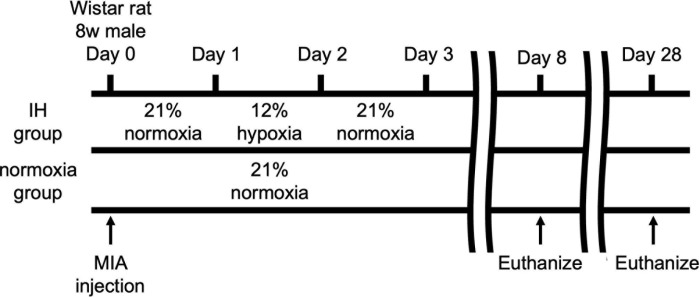
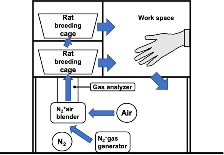
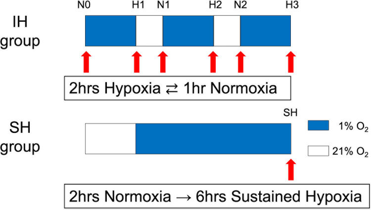
Hypoxia-inducible factor-1α plays an important role in the homeostasis of articular cartilage in hypoxic environments. Therefore, modulation of hypoxia-inducible factor-1α by regulating the oxygen environment could be a useful treatment for osteoarthritis. This study aimed to assess the chondroprotective effects of intermittent hypoxia on cultured chondrocytes and an animal model of osteoarthritis. In vitro, human chondrocytes were exposed to 2 h of hypoxic stimulation three times at 1-h intervals, and protein and gene expression of hypoxia-inducible factor-1α, ACAN, and cell viability was measured over time. In vivo, 8-week-old male Wistar rats were injected with monosodium iodoacetate to induce osteoarthritis and then reared in 12% hypoxia for 24 h, followed by 24 h in steady oxygen, repeated alternately for a total of 28 days. A histological analysis was performed on days 8 and 28. In the intermittent hypoxia group, each protein expression increased with each repeated hypoxic stimulation to human chondrocytes; finally, the protein level was significantly higher with intermittent hypoxia than with continuous hypoxic stimulation, cell viability was increased, and gene expression was not significantly increased. In the osteoarthritis animal model, for 8 days, there were stronger hypoxia-inducible factor-1α staining and no significant differences in articular cartilage destruction. Furthermore, for 28 days, there was significantly less articular cartilage destruction in the rat osteoarthritis model with intermittent hypoxia than with steady oxygen rearing. Intermittent hypoxia increased cartilage metabolism by increasing hypoxia-inducible factor-1α proteins in articular chondrocytes, which may be effective in preventing articular cartilage degeneration in a rat osteoarthritis model.
期刊介绍:
PLOS ONE is an international, peer-reviewed, open-access, online publication. PLOS ONE welcomes reports on primary research from any scientific discipline. It provides:
* Open-access—freely accessible online, authors retain copyright
* Fast publication times
* Peer review by expert, practicing researchers
* Post-publication tools to indicate quality and impact
* Community-based dialogue on articles
* Worldwide media coverage

 求助内容:
求助内容: 应助结果提醒方式:


