{"title":"抗vegf治疗后黄斑水肿复发与黄斑结构及血管特征的关系","authors":"Dazhuang Ren, Cece Zhao, Gaoxu Wei, Xiaoyun Hou, Chang Li, Zhiqing Li","doi":"10.1155/joph/8824342","DOIUrl":null,"url":null,"abstract":"<p><p><b>Purpose:</b> To identify the structural and vascular features of the macula related to the recurrence of macular edema (ME) in central retinal vein occlusion (CRVO) after intravitreal anti-VEGF injections. <b>Methods:</b> This was a cross-sectional study including CRVO patients without ME and age-matched individuals. CRVO patients were divided into the ME-resolved group and the ME-recurrence group on the basis of whether ME recurred within 3 months. All subjects provided a detailed history and underwent a comprehensive ophthalmological examination. Measurements of the macula by swept-source optical coherence tomography angiography (SS-OCTA) were recorded. We also created the Δparameter, which represents the difference in OCTA parameters between CRVO-affected eyes and their fellow eyes. <b>Results:</b> The study included 13 ME-resolved CRVO patients, 20 ME-recurrence CRVO patients, and 24 age-matched controls. Compared with the ME-recurrence group, the ME-resolved group had a longer CRVO duration, more previous intravitreal anti-VEGF injections, and a higher proportion of previous retinal photocoagulation (all <i>p</i> < 0.05). Additionally, retinal thickness (RT) and choroidal thickness (CT) were thinner in the ME-resolved group than in the ME-recurrence and control groups (all <i>p</i> < 0.01). The ME-resolved group also had significantly lower vessel density (VD) in both superficial and deep vascular complexes (SVC/DVC) and larger foveal avascular zone area (FAZa) in SVC and DVC than the ME-recurrence group and the control group (all <i>p</i> < 0.01). The results were the same with the Δparameters. Multivariate logistic regression revealed that ΔVD and ΔFAZa in SVC and DVC were independently associated with ME recurrence after adjusting for the effects of CRVO duration, previous anti-VEGF injections, and retinal photocoagulation (all <i>p</i> < 0.05). <b>Conclusion:</b> With prolonged CRVO duration, more anti-VEGF injections, and more retinal photocoagulation procedures, retinal, choroidal, and vascular atrophy in the macula occurs in CRVO eyes, making ME less likely to recur. Macular vascular atrophy is vital for the resolution of ME and might be a manifestation of capillary remodeling.</p>","PeriodicalId":16674,"journal":{"name":"Journal of Ophthalmology","volume":"2025 ","pages":"8824342"},"PeriodicalIF":1.9000,"publicationDate":"2025-02-28","publicationTypes":"Journal Article","fieldsOfStudy":null,"isOpenAccess":false,"openAccessPdf":"https://www.ncbi.nlm.nih.gov/pmc/articles/PMC11961281/pdf/","citationCount":"0","resultStr":"{\"title\":\"Structural and Vascular Features of Macula Related to the Recurrence of Macular Edema in Central Retinal Vein Occlusion After Anti-VEGF Therapy.\",\"authors\":\"Dazhuang Ren, Cece Zhao, Gaoxu Wei, Xiaoyun Hou, Chang Li, Zhiqing Li\",\"doi\":\"10.1155/joph/8824342\",\"DOIUrl\":null,\"url\":null,\"abstract\":\"<p><p><b>Purpose:</b> To identify the structural and vascular features of the macula related to the recurrence of macular edema (ME) in central retinal vein occlusion (CRVO) after intravitreal anti-VEGF injections. <b>Methods:</b> This was a cross-sectional study including CRVO patients without ME and age-matched individuals. CRVO patients were divided into the ME-resolved group and the ME-recurrence group on the basis of whether ME recurred within 3 months. All subjects provided a detailed history and underwent a comprehensive ophthalmological examination. Measurements of the macula by swept-source optical coherence tomography angiography (SS-OCTA) were recorded. We also created the Δparameter, which represents the difference in OCTA parameters between CRVO-affected eyes and their fellow eyes. <b>Results:</b> The study included 13 ME-resolved CRVO patients, 20 ME-recurrence CRVO patients, and 24 age-matched controls. Compared with the ME-recurrence group, the ME-resolved group had a longer CRVO duration, more previous intravitreal anti-VEGF injections, and a higher proportion of previous retinal photocoagulation (all <i>p</i> < 0.05). Additionally, retinal thickness (RT) and choroidal thickness (CT) were thinner in the ME-resolved group than in the ME-recurrence and control groups (all <i>p</i> < 0.01). The ME-resolved group also had significantly lower vessel density (VD) in both superficial and deep vascular complexes (SVC/DVC) and larger foveal avascular zone area (FAZa) in SVC and DVC than the ME-recurrence group and the control group (all <i>p</i> < 0.01). The results were the same with the Δparameters. Multivariate logistic regression revealed that ΔVD and ΔFAZa in SVC and DVC were independently associated with ME recurrence after adjusting for the effects of CRVO duration, previous anti-VEGF injections, and retinal photocoagulation (all <i>p</i> < 0.05). <b>Conclusion:</b> With prolonged CRVO duration, more anti-VEGF injections, and more retinal photocoagulation procedures, retinal, choroidal, and vascular atrophy in the macula occurs in CRVO eyes, making ME less likely to recur. Macular vascular atrophy is vital for the resolution of ME and might be a manifestation of capillary remodeling.</p>\",\"PeriodicalId\":16674,\"journal\":{\"name\":\"Journal of Ophthalmology\",\"volume\":\"2025 \",\"pages\":\"8824342\"},\"PeriodicalIF\":1.9000,\"publicationDate\":\"2025-02-28\",\"publicationTypes\":\"Journal Article\",\"fieldsOfStudy\":null,\"isOpenAccess\":false,\"openAccessPdf\":\"https://www.ncbi.nlm.nih.gov/pmc/articles/PMC11961281/pdf/\",\"citationCount\":\"0\",\"resultStr\":null,\"platform\":\"Semanticscholar\",\"paperid\":null,\"PeriodicalName\":\"Journal of Ophthalmology\",\"FirstCategoryId\":\"3\",\"ListUrlMain\":\"https://doi.org/10.1155/joph/8824342\",\"RegionNum\":4,\"RegionCategory\":\"医学\",\"ArticlePicture\":[],\"TitleCN\":null,\"AbstractTextCN\":null,\"PMCID\":null,\"EPubDate\":\"2025/1/1 0:00:00\",\"PubModel\":\"eCollection\",\"JCR\":\"Q3\",\"JCRName\":\"OPHTHALMOLOGY\",\"Score\":null,\"Total\":0}","platform":"Semanticscholar","paperid":null,"PeriodicalName":"Journal of Ophthalmology","FirstCategoryId":"3","ListUrlMain":"https://doi.org/10.1155/joph/8824342","RegionNum":4,"RegionCategory":"医学","ArticlePicture":[],"TitleCN":null,"AbstractTextCN":null,"PMCID":null,"EPubDate":"2025/1/1 0:00:00","PubModel":"eCollection","JCR":"Q3","JCRName":"OPHTHALMOLOGY","Score":null,"Total":0}
Structural and Vascular Features of Macula Related to the Recurrence of Macular Edema in Central Retinal Vein Occlusion After Anti-VEGF Therapy.
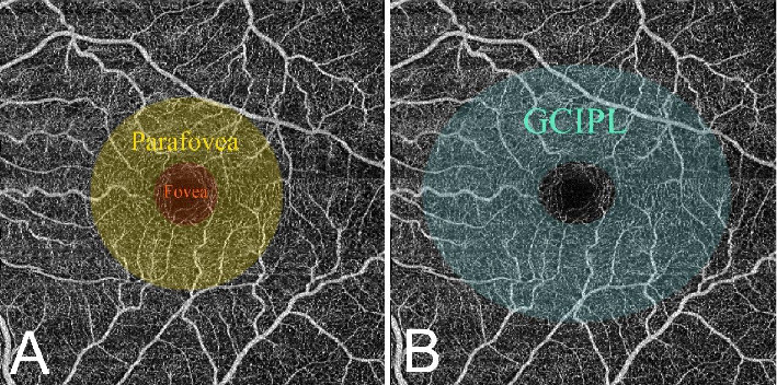
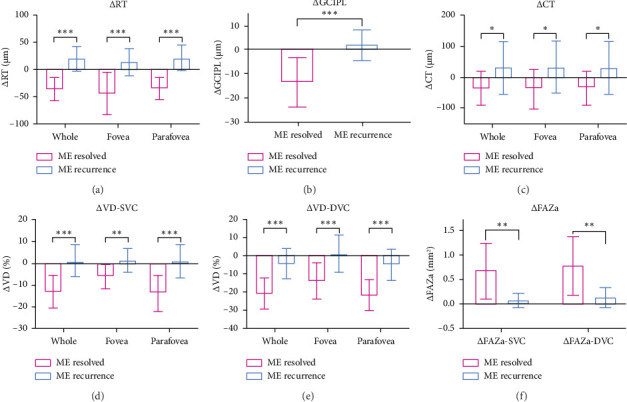
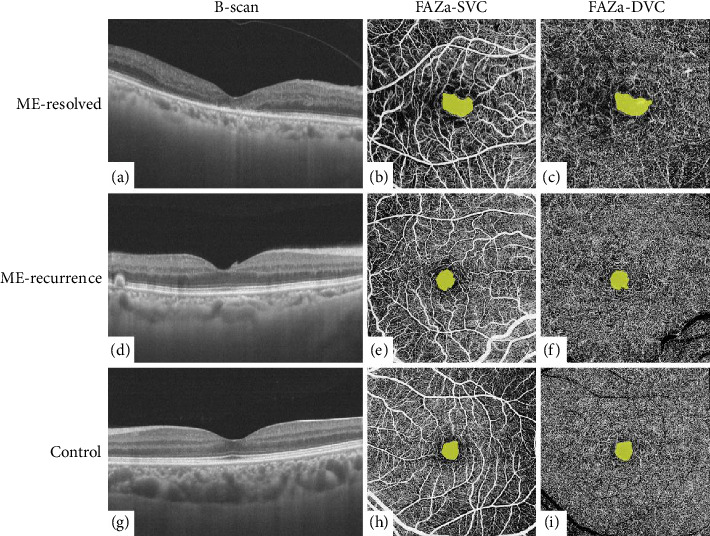
Purpose: To identify the structural and vascular features of the macula related to the recurrence of macular edema (ME) in central retinal vein occlusion (CRVO) after intravitreal anti-VEGF injections. Methods: This was a cross-sectional study including CRVO patients without ME and age-matched individuals. CRVO patients were divided into the ME-resolved group and the ME-recurrence group on the basis of whether ME recurred within 3 months. All subjects provided a detailed history and underwent a comprehensive ophthalmological examination. Measurements of the macula by swept-source optical coherence tomography angiography (SS-OCTA) were recorded. We also created the Δparameter, which represents the difference in OCTA parameters between CRVO-affected eyes and their fellow eyes. Results: The study included 13 ME-resolved CRVO patients, 20 ME-recurrence CRVO patients, and 24 age-matched controls. Compared with the ME-recurrence group, the ME-resolved group had a longer CRVO duration, more previous intravitreal anti-VEGF injections, and a higher proportion of previous retinal photocoagulation (all p < 0.05). Additionally, retinal thickness (RT) and choroidal thickness (CT) were thinner in the ME-resolved group than in the ME-recurrence and control groups (all p < 0.01). The ME-resolved group also had significantly lower vessel density (VD) in both superficial and deep vascular complexes (SVC/DVC) and larger foveal avascular zone area (FAZa) in SVC and DVC than the ME-recurrence group and the control group (all p < 0.01). The results were the same with the Δparameters. Multivariate logistic regression revealed that ΔVD and ΔFAZa in SVC and DVC were independently associated with ME recurrence after adjusting for the effects of CRVO duration, previous anti-VEGF injections, and retinal photocoagulation (all p < 0.05). Conclusion: With prolonged CRVO duration, more anti-VEGF injections, and more retinal photocoagulation procedures, retinal, choroidal, and vascular atrophy in the macula occurs in CRVO eyes, making ME less likely to recur. Macular vascular atrophy is vital for the resolution of ME and might be a manifestation of capillary remodeling.
期刊介绍:
Journal of Ophthalmology is a peer-reviewed, Open Access journal that publishes original research articles, review articles, and clinical studies related to the anatomy, physiology and diseases of the eye. Submissions should focus on new diagnostic and surgical techniques, instrument and therapy updates, as well as clinical trials and research findings.
 求助内容:
求助内容: 应助结果提醒方式:


