{"title":"子宫内膜病变患者液基细胞学准备中细胞的形态特征和排列。","authors":"Ming-Zhe Wu, Na-Jin Gu, Ming-Ming Xiao, Dong-Ge Liu, Mu-Lan Jin, Xu-Yan Liu, Jian Wang, Hong-Tao Xu, Yi Zhang, Guang-Ping Wu","doi":"10.1186/s13000-025-01631-4","DOIUrl":null,"url":null,"abstract":"<p><strong>Background: </strong>The accurate cytological diagnosis of endometrial carcinomas by minimally invasive method has a broad application. There are several articles described the morphological characteristics but not arrangements of endometrial lesion cells on LBC slides.</p><p><strong>Methods: </strong>A retrospective study was conducted using 175 endometrial samples obtained by direct negative pressure suction with disposable endometrial sampler. All lesions were diagnosed both cytologically and histologically, and the diagnostic results were compared and analyzed.</p><p><strong>Results: </strong>The cytological diagnoses of polyps, simple or complex hyperplasia, and atypical hyperplasia were highly consistent with the histological diagnosis. The cytological features of polyps and normal endometrium, as well as simple and complex hyperplasia, are the same. Among 82 cases of histologically confirmed adenocarcinoma, the cytological diagnosis were adenocarcinoma cells (46 cases, 56.10%), suspected for adenocarcinoma cells (22 cases, 26.83%), and false negative (14 cases,17.07%). Retrospective reviewing the slide suggest diagnostic parameters such as significantly enlarged nuclei, multistage papillary arrangements, large and numerous nucleoli, and large vacuoles containing neutrophils in the cytoplasm are reliable diagnostic criteria for endometrial carcinoma cells; on the other hand, ignorance of lobulated arrangements and escaped arrangements are the main reasons for missed diagnosis.</p><p><strong>Conclusions: </strong>The cytological diagnosis of endometrial lesions not only depends on the morphological characteristics of cells, but also need careful observations of the cellular arrangements.</p>","PeriodicalId":11237,"journal":{"name":"Diagnostic Pathology","volume":"20 1","pages":"32"},"PeriodicalIF":2.3000,"publicationDate":"2025-04-01","publicationTypes":"Journal Article","fieldsOfStudy":null,"isOpenAccess":false,"openAccessPdf":"https://www.ncbi.nlm.nih.gov/pmc/articles/PMC11963423/pdf/","citationCount":"0","resultStr":"{\"title\":\"The morphological characteristics and arrangements of cells in the liquid-based cytology preparation of patients with endometrial lesions.\",\"authors\":\"Ming-Zhe Wu, Na-Jin Gu, Ming-Ming Xiao, Dong-Ge Liu, Mu-Lan Jin, Xu-Yan Liu, Jian Wang, Hong-Tao Xu, Yi Zhang, Guang-Ping Wu\",\"doi\":\"10.1186/s13000-025-01631-4\",\"DOIUrl\":null,\"url\":null,\"abstract\":\"<p><strong>Background: </strong>The accurate cytological diagnosis of endometrial carcinomas by minimally invasive method has a broad application. There are several articles described the morphological characteristics but not arrangements of endometrial lesion cells on LBC slides.</p><p><strong>Methods: </strong>A retrospective study was conducted using 175 endometrial samples obtained by direct negative pressure suction with disposable endometrial sampler. All lesions were diagnosed both cytologically and histologically, and the diagnostic results were compared and analyzed.</p><p><strong>Results: </strong>The cytological diagnoses of polyps, simple or complex hyperplasia, and atypical hyperplasia were highly consistent with the histological diagnosis. The cytological features of polyps and normal endometrium, as well as simple and complex hyperplasia, are the same. Among 82 cases of histologically confirmed adenocarcinoma, the cytological diagnosis were adenocarcinoma cells (46 cases, 56.10%), suspected for adenocarcinoma cells (22 cases, 26.83%), and false negative (14 cases,17.07%). Retrospective reviewing the slide suggest diagnostic parameters such as significantly enlarged nuclei, multistage papillary arrangements, large and numerous nucleoli, and large vacuoles containing neutrophils in the cytoplasm are reliable diagnostic criteria for endometrial carcinoma cells; on the other hand, ignorance of lobulated arrangements and escaped arrangements are the main reasons for missed diagnosis.</p><p><strong>Conclusions: </strong>The cytological diagnosis of endometrial lesions not only depends on the morphological characteristics of cells, but also need careful observations of the cellular arrangements.</p>\",\"PeriodicalId\":11237,\"journal\":{\"name\":\"Diagnostic Pathology\",\"volume\":\"20 1\",\"pages\":\"32\"},\"PeriodicalIF\":2.3000,\"publicationDate\":\"2025-04-01\",\"publicationTypes\":\"Journal Article\",\"fieldsOfStudy\":null,\"isOpenAccess\":false,\"openAccessPdf\":\"https://www.ncbi.nlm.nih.gov/pmc/articles/PMC11963423/pdf/\",\"citationCount\":\"0\",\"resultStr\":null,\"platform\":\"Semanticscholar\",\"paperid\":null,\"PeriodicalName\":\"Diagnostic Pathology\",\"FirstCategoryId\":\"3\",\"ListUrlMain\":\"https://doi.org/10.1186/s13000-025-01631-4\",\"RegionNum\":3,\"RegionCategory\":\"医学\",\"ArticlePicture\":[],\"TitleCN\":null,\"AbstractTextCN\":null,\"PMCID\":null,\"EPubDate\":\"\",\"PubModel\":\"\",\"JCR\":\"Q2\",\"JCRName\":\"PATHOLOGY\",\"Score\":null,\"Total\":0}","platform":"Semanticscholar","paperid":null,"PeriodicalName":"Diagnostic Pathology","FirstCategoryId":"3","ListUrlMain":"https://doi.org/10.1186/s13000-025-01631-4","RegionNum":3,"RegionCategory":"医学","ArticlePicture":[],"TitleCN":null,"AbstractTextCN":null,"PMCID":null,"EPubDate":"","PubModel":"","JCR":"Q2","JCRName":"PATHOLOGY","Score":null,"Total":0}
The morphological characteristics and arrangements of cells in the liquid-based cytology preparation of patients with endometrial lesions.
Background: The accurate cytological diagnosis of endometrial carcinomas by minimally invasive method has a broad application. There are several articles described the morphological characteristics but not arrangements of endometrial lesion cells on LBC slides.
Methods: A retrospective study was conducted using 175 endometrial samples obtained by direct negative pressure suction with disposable endometrial sampler. All lesions were diagnosed both cytologically and histologically, and the diagnostic results were compared and analyzed.
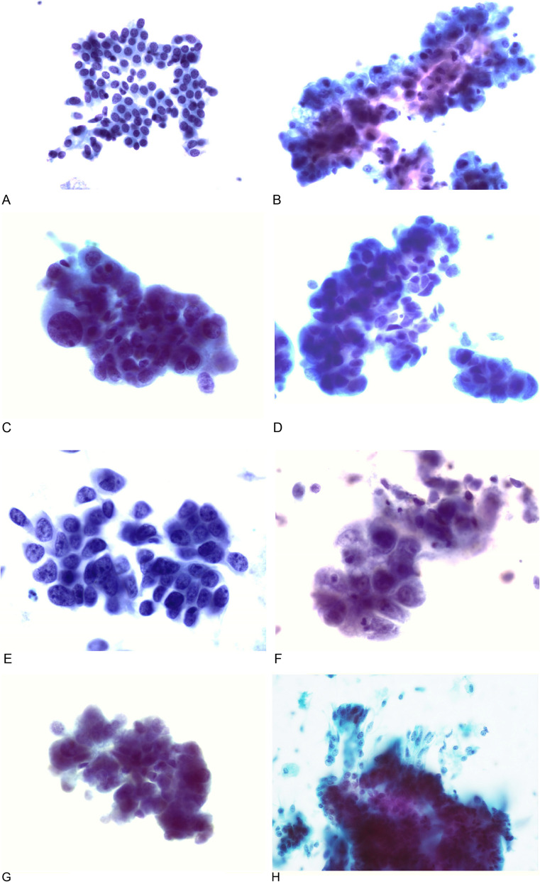
Results: The cytological diagnoses of polyps, simple or complex hyperplasia, and atypical hyperplasia were highly consistent with the histological diagnosis. The cytological features of polyps and normal endometrium, as well as simple and complex hyperplasia, are the same. Among 82 cases of histologically confirmed adenocarcinoma, the cytological diagnosis were adenocarcinoma cells (46 cases, 56.10%), suspected for adenocarcinoma cells (22 cases, 26.83%), and false negative (14 cases,17.07%). Retrospective reviewing the slide suggest diagnostic parameters such as significantly enlarged nuclei, multistage papillary arrangements, large and numerous nucleoli, and large vacuoles containing neutrophils in the cytoplasm are reliable diagnostic criteria for endometrial carcinoma cells; on the other hand, ignorance of lobulated arrangements and escaped arrangements are the main reasons for missed diagnosis.
Conclusions: The cytological diagnosis of endometrial lesions not only depends on the morphological characteristics of cells, but also need careful observations of the cellular arrangements.
期刊介绍:
Diagnostic Pathology is an open access, peer-reviewed, online journal that considers research in surgical and clinical pathology, immunology, and biology, with a special focus on cutting-edge approaches in diagnostic pathology and tissue-based therapy. The journal covers all aspects of surgical pathology, including classic diagnostic pathology, prognosis-related diagnosis (tumor stages, prognosis markers, such as MIB-percentage, hormone receptors, etc.), and therapy-related findings. The journal also focuses on the technological aspects of pathology, including molecular biology techniques, morphometry aspects (stereology, DNA analysis, syntactic structure analysis), communication aspects (telecommunication, virtual microscopy, virtual pathology institutions, etc.), and electronic education and quality assurance (for example interactive publication, on-line references with automated updating, etc.).

 求助内容:
求助内容: 应助结果提醒方式:


