{"title":"睾丸横向异位合并持续性<s:1>勒管综合征1例。","authors":"Keisuke Hidaka, Akihiro Furuta, Nobuyuki Mori, Takuya Maekura, Fuki Shitano, Yang Wang, Naoko Nishio, Yumiko Fujiwara, Toshiya Takamura, Kotoe Hidaka, Toshiki Shiozaki","doi":"10.1007/s00261-025-04915-x","DOIUrl":null,"url":null,"abstract":"<div><p>Transverse testicular ectopia (TTE) is a variant of ectopic testis in which both testes are located in the same scrotum. Persistent Müllerian duct syndrome (PMDS) is a congenital anomaly characterized by the retention of Müllerian duct remnants in males. TTE is a characteristic clinical finding in PMDS. This case report describes a patient in his 50s who was diagnosed with PMDS based on TTE findings and a pelvic structure suggestive of a Müllerian duct remnant. Computed tomography (CT) and magnetic resonance imaging (MRI) revealed the normal left testis within the left scrotum. The soft tissue was identified dorsal to the left testis, supplied from branches of bilateral internal iliac arteries. Additionally, a rod-like structure was observed in the left pelvis, appearing continuous with the soft tissue in the left scrotum. The right testis, enlarged and torsed, was located cranially within the left scrotum. A cord-like structure extended continuously from this ectopically positioned right testis toward the right inguinal region. Surgical findings revealed a normal left testis and torsion of the right testis. The right testis was removed. Histological examination revealed a seminoma within the right testis, and immunostaining confirmed the presence of attached Müllerian duct remnants. Prior imaging revealed that the right testis was located in the abdominal cavity, indicating that TTE in this case did not occur during testicular descent. In PMDS cases, TTE is believed to result from the traction of one testis by Müllerian duct remnants and the testicular mobility due to an abnormally long gubernaculum, which is a crucial role in normal testicular descent. The findings in this case support this theory and further indicate that TTE can occur even after testicular descent is complete. This report describes a case of TTE with a causative Müllerian duct remnant and a presumed abnormally long gubernaculum identified on imaging, which contributes to the understanding of this rare condition.</p><h3>Graphical abstract</h3><div><figure><div><div><picture><source><img></source></picture></div></div></figure></div></div>","PeriodicalId":7126,"journal":{"name":"Abdominal Radiology","volume":"50 10","pages":"4796 - 4802"},"PeriodicalIF":2.2000,"publicationDate":"2025-04-02","publicationTypes":"Journal Article","fieldsOfStudy":null,"isOpenAccess":false,"openAccessPdf":"","citationCount":"0","resultStr":"{\"title\":\"Transverse testicular ectopia with persistent müllerian duct syndrome: a case report\",\"authors\":\"Keisuke Hidaka, Akihiro Furuta, Nobuyuki Mori, Takuya Maekura, Fuki Shitano, Yang Wang, Naoko Nishio, Yumiko Fujiwara, Toshiya Takamura, Kotoe Hidaka, Toshiki Shiozaki\",\"doi\":\"10.1007/s00261-025-04915-x\",\"DOIUrl\":null,\"url\":null,\"abstract\":\"<div><p>Transverse testicular ectopia (TTE) is a variant of ectopic testis in which both testes are located in the same scrotum. Persistent Müllerian duct syndrome (PMDS) is a congenital anomaly characterized by the retention of Müllerian duct remnants in males. TTE is a characteristic clinical finding in PMDS. This case report describes a patient in his 50s who was diagnosed with PMDS based on TTE findings and a pelvic structure suggestive of a Müllerian duct remnant. Computed tomography (CT) and magnetic resonance imaging (MRI) revealed the normal left testis within the left scrotum. The soft tissue was identified dorsal to the left testis, supplied from branches of bilateral internal iliac arteries. Additionally, a rod-like structure was observed in the left pelvis, appearing continuous with the soft tissue in the left scrotum. The right testis, enlarged and torsed, was located cranially within the left scrotum. A cord-like structure extended continuously from this ectopically positioned right testis toward the right inguinal region. Surgical findings revealed a normal left testis and torsion of the right testis. The right testis was removed. Histological examination revealed a seminoma within the right testis, and immunostaining confirmed the presence of attached Müllerian duct remnants. Prior imaging revealed that the right testis was located in the abdominal cavity, indicating that TTE in this case did not occur during testicular descent. In PMDS cases, TTE is believed to result from the traction of one testis by Müllerian duct remnants and the testicular mobility due to an abnormally long gubernaculum, which is a crucial role in normal testicular descent. The findings in this case support this theory and further indicate that TTE can occur even after testicular descent is complete. This report describes a case of TTE with a causative Müllerian duct remnant and a presumed abnormally long gubernaculum identified on imaging, which contributes to the understanding of this rare condition.</p><h3>Graphical abstract</h3><div><figure><div><div><picture><source><img></source></picture></div></div></figure></div></div>\",\"PeriodicalId\":7126,\"journal\":{\"name\":\"Abdominal Radiology\",\"volume\":\"50 10\",\"pages\":\"4796 - 4802\"},\"PeriodicalIF\":2.2000,\"publicationDate\":\"2025-04-02\",\"publicationTypes\":\"Journal Article\",\"fieldsOfStudy\":null,\"isOpenAccess\":false,\"openAccessPdf\":\"\",\"citationCount\":\"0\",\"resultStr\":null,\"platform\":\"Semanticscholar\",\"paperid\":null,\"PeriodicalName\":\"Abdominal Radiology\",\"FirstCategoryId\":\"3\",\"ListUrlMain\":\"https://link.springer.com/article/10.1007/s00261-025-04915-x\",\"RegionNum\":3,\"RegionCategory\":\"医学\",\"ArticlePicture\":[],\"TitleCN\":null,\"AbstractTextCN\":null,\"PMCID\":null,\"EPubDate\":\"\",\"PubModel\":\"\",\"JCR\":\"Q2\",\"JCRName\":\"RADIOLOGY, NUCLEAR MEDICINE & MEDICAL IMAGING\",\"Score\":null,\"Total\":0}","platform":"Semanticscholar","paperid":null,"PeriodicalName":"Abdominal Radiology","FirstCategoryId":"3","ListUrlMain":"https://link.springer.com/article/10.1007/s00261-025-04915-x","RegionNum":3,"RegionCategory":"医学","ArticlePicture":[],"TitleCN":null,"AbstractTextCN":null,"PMCID":null,"EPubDate":"","PubModel":"","JCR":"Q2","JCRName":"RADIOLOGY, NUCLEAR MEDICINE & MEDICAL IMAGING","Score":null,"Total":0}
Transverse testicular ectopia with persistent müllerian duct syndrome: a case report
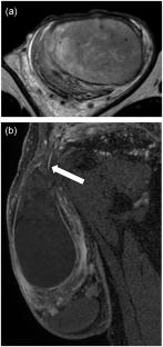
Transverse testicular ectopia (TTE) is a variant of ectopic testis in which both testes are located in the same scrotum. Persistent Müllerian duct syndrome (PMDS) is a congenital anomaly characterized by the retention of Müllerian duct remnants in males. TTE is a characteristic clinical finding in PMDS. This case report describes a patient in his 50s who was diagnosed with PMDS based on TTE findings and a pelvic structure suggestive of a Müllerian duct remnant. Computed tomography (CT) and magnetic resonance imaging (MRI) revealed the normal left testis within the left scrotum. The soft tissue was identified dorsal to the left testis, supplied from branches of bilateral internal iliac arteries. Additionally, a rod-like structure was observed in the left pelvis, appearing continuous with the soft tissue in the left scrotum. The right testis, enlarged and torsed, was located cranially within the left scrotum. A cord-like structure extended continuously from this ectopically positioned right testis toward the right inguinal region. Surgical findings revealed a normal left testis and torsion of the right testis. The right testis was removed. Histological examination revealed a seminoma within the right testis, and immunostaining confirmed the presence of attached Müllerian duct remnants. Prior imaging revealed that the right testis was located in the abdominal cavity, indicating that TTE in this case did not occur during testicular descent. In PMDS cases, TTE is believed to result from the traction of one testis by Müllerian duct remnants and the testicular mobility due to an abnormally long gubernaculum, which is a crucial role in normal testicular descent. The findings in this case support this theory and further indicate that TTE can occur even after testicular descent is complete. This report describes a case of TTE with a causative Müllerian duct remnant and a presumed abnormally long gubernaculum identified on imaging, which contributes to the understanding of this rare condition.
期刊介绍:
Abdominal Radiology seeks to meet the professional needs of the abdominal radiologist by publishing clinically pertinent original, review and practice related articles on the gastrointestinal and genitourinary tracts and abdominal interventional and radiologic procedures. Case reports are generally not accepted unless they are the first report of a new disease or condition, or part of a special solicited section.
Reasons to Publish Your Article in Abdominal Radiology:
· Official journal of the Society of Abdominal Radiology (SAR)
· Published in Cooperation with:
European Society of Gastrointestinal and Abdominal Radiology (ESGAR)
European Society of Urogenital Radiology (ESUR)
Asian Society of Abdominal Radiology (ASAR)
· Efficient handling and Expeditious review
· Author feedback is provided in a mentoring style
· Global readership
· Readers can earn CME credits


 求助内容:
求助内容: 应助结果提醒方式:


