3D打印骨质疏松症骨模型在动态培养中得到验证
Q1 Computer Science
引用次数: 0
摘要
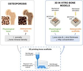
骨质疏松症是一种世界性的骨病,其特征是骨量减少和骨结构改变,导致骨脆性和骨折风险增加。尽管动物模型仍然是研究和测试新型抗骨质疏松药物的金标准,但动物模型价格昂贵且无法准确再现体内条件,因此迫切需要用替代方法替代动物模型。在骨组织工程领域,病理三维(3D)体外骨模型最近被认为克服了与传统临床前测试方法相关的经济和伦理问题。因此,本研究旨在设计一个骨质疏松性骨的3D体外模型,该模型由3D打印支架组成,该支架与生理性骨和骨质疏松性骨的结构和骨矿物质含量差异相似,并将成骨前细胞植入支架上。设计并打印体外生理三维骨模型作为对照条件。在扫描电镜下对未播种的支架进行了全面的物理化学表征,包括机械和热性能、膨胀行为、降解和形态学检查。在机械刺激下培养细胞种子生理性和骨质疏松性骨支架,以模拟人类骨骼日常承受的机械力。机械刺激的应用对成骨前细胞的成骨分化有显著的积极影响,其中细胞种子的骨质疏松支架的成骨分化值最低,因此类似于骨质疏松患者骨形成的减少。本文章由计算机程序翻译,如有差异,请以英文原文为准。
3D printed osteoporotic bone model validated in dynamic culture
Osteoporosis is a worldwide bone disease characterized by reduced bone mass and an alteration of bone architecture, leading to bone fragility and an increased risk of fractures. Although animal models are still the gold standard for studying and testing new anti-osteoporotic drugs, they are expensive and unable to reproduce the in vivo conditions accurately, thus making their replacement with alternative methods an urgent need. In the field of bone tissue engineering, pathological three-dimensional (3D) in vitro bone models have been recently considered to overcome economic and ethical issues associated with traditional pre-clinical testing methods. As a result, this study aimed to design a 3D in vitro model of osteoporotic bone consisting of 3D printed scaffolds that resemble the architectural and bone mineral content differences between physiological and osteoporotic bone, and pre-osteoblastic cells seeded onto the scaffolds. A physiological 3D in vitro bone model was designed and printed as a control condition. A comprehensive physicochemical characterization of unseeded scaffolds was conducted in terms of mechanical and thermal properties, swelling behaviour, degradation, and morphology examination under scanning electron microscopy. Cell-seeded physiological and osteoporotic bone scaffolds were cultured under mechanical stimulation to mimic the mechanical forces experienced daily by human bones. The application of mechanical stimuli had a significantly positive effect on the osteogenic differentiation of the pre-osteoblastic cells, with cell-seeded osteoporotic scaffolds reporting the lowest values, thus resembling the reduction in bone formation in osteoporotic patients.
求助全文
通过发布文献求助,成功后即可免费获取论文全文。
去求助
来源期刊

Bioprinting
Computer Science-Computer Science Applications
CiteScore
11.50
自引率
0.00%
发文量
72
审稿时长
68 days
期刊介绍:
Bioprinting is a broad-spectrum, multidisciplinary journal that covers all aspects of 3D fabrication technology involving biological tissues, organs and cells for medical and biotechnology applications. Topics covered include nanomaterials, biomaterials, scaffolds, 3D printing technology, imaging and CAD/CAM software and hardware, post-printing bioreactor maturation, cell and biological factor patterning, biofabrication, tissue engineering and other applications of 3D bioprinting technology. Bioprinting publishes research reports describing novel results with high clinical significance in all areas of 3D bioprinting research. Bioprinting issues contain a wide variety of review and analysis articles covering topics relevant to 3D bioprinting ranging from basic biological, material and technical advances to pre-clinical and clinical applications of 3D bioprinting.
 求助内容:
求助内容: 应助结果提醒方式:


