{"title":"猫双肠套叠的超声特征。","authors":"Stefano Ludovici, Anna Cronin, Domenico Sainato","doi":"10.1177/20551169251316999","DOIUrl":null,"url":null,"abstract":"<p><strong>Case summary: </strong>A 6-year-old male castrated Maine Coon cat was presented with a 3-day history of lethargy, hyporexia and weight loss. Abdominal ultrasonography demonstrated a double intestinal intussusception with the colon intussuscepting a thickened ileal segment, which was in turn intussuscepting the jejunum. A jejunal prolapse through the anus occurred 3 days later as a complication of the double intussusception, at which time the cat underwent surgery. Manual reduction of part of the intussusception was achieved, while the remaining 30 cm, including of the ileocaecocolic junction, was resected followed by functional end-to-end anastomosis. The cat recovered uneventfully without any reported long-term gastrointestinal complications.</p><p><strong>Relevance and novel information: </strong>Double intussusception is rare in cats. To the best of the authors' knowledge, this is the first case to describe ultrasonographic features of double intussusception in a cat.</p>","PeriodicalId":36588,"journal":{"name":"Journal of Feline Medicine and Surgery Open Reports","volume":"11 1","pages":"20551169251316999"},"PeriodicalIF":0.7000,"publicationDate":"2025-03-28","publicationTypes":"Journal Article","fieldsOfStudy":null,"isOpenAccess":false,"openAccessPdf":"https://www.ncbi.nlm.nih.gov/pmc/articles/PMC11954383/pdf/","citationCount":"0","resultStr":"{\"title\":\"Ultrasonographic features of double intestinal intussusception in a cat.\",\"authors\":\"Stefano Ludovici, Anna Cronin, Domenico Sainato\",\"doi\":\"10.1177/20551169251316999\",\"DOIUrl\":null,\"url\":null,\"abstract\":\"<p><strong>Case summary: </strong>A 6-year-old male castrated Maine Coon cat was presented with a 3-day history of lethargy, hyporexia and weight loss. Abdominal ultrasonography demonstrated a double intestinal intussusception with the colon intussuscepting a thickened ileal segment, which was in turn intussuscepting the jejunum. A jejunal prolapse through the anus occurred 3 days later as a complication of the double intussusception, at which time the cat underwent surgery. Manual reduction of part of the intussusception was achieved, while the remaining 30 cm, including of the ileocaecocolic junction, was resected followed by functional end-to-end anastomosis. The cat recovered uneventfully without any reported long-term gastrointestinal complications.</p><p><strong>Relevance and novel information: </strong>Double intussusception is rare in cats. To the best of the authors' knowledge, this is the first case to describe ultrasonographic features of double intussusception in a cat.</p>\",\"PeriodicalId\":36588,\"journal\":{\"name\":\"Journal of Feline Medicine and Surgery Open Reports\",\"volume\":\"11 1\",\"pages\":\"20551169251316999\"},\"PeriodicalIF\":0.7000,\"publicationDate\":\"2025-03-28\",\"publicationTypes\":\"Journal Article\",\"fieldsOfStudy\":null,\"isOpenAccess\":false,\"openAccessPdf\":\"https://www.ncbi.nlm.nih.gov/pmc/articles/PMC11954383/pdf/\",\"citationCount\":\"0\",\"resultStr\":null,\"platform\":\"Semanticscholar\",\"paperid\":null,\"PeriodicalName\":\"Journal of Feline Medicine and Surgery Open Reports\",\"FirstCategoryId\":\"1085\",\"ListUrlMain\":\"https://doi.org/10.1177/20551169251316999\",\"RegionNum\":0,\"RegionCategory\":null,\"ArticlePicture\":[],\"TitleCN\":null,\"AbstractTextCN\":null,\"PMCID\":null,\"EPubDate\":\"2025/1/1 0:00:00\",\"PubModel\":\"eCollection\",\"JCR\":\"Q3\",\"JCRName\":\"VETERINARY SCIENCES\",\"Score\":null,\"Total\":0}","platform":"Semanticscholar","paperid":null,"PeriodicalName":"Journal of Feline Medicine and Surgery Open Reports","FirstCategoryId":"1085","ListUrlMain":"https://doi.org/10.1177/20551169251316999","RegionNum":0,"RegionCategory":null,"ArticlePicture":[],"TitleCN":null,"AbstractTextCN":null,"PMCID":null,"EPubDate":"2025/1/1 0:00:00","PubModel":"eCollection","JCR":"Q3","JCRName":"VETERINARY SCIENCES","Score":null,"Total":0}
Ultrasonographic features of double intestinal intussusception in a cat.
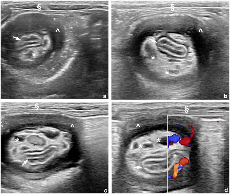
Case summary: A 6-year-old male castrated Maine Coon cat was presented with a 3-day history of lethargy, hyporexia and weight loss. Abdominal ultrasonography demonstrated a double intestinal intussusception with the colon intussuscepting a thickened ileal segment, which was in turn intussuscepting the jejunum. A jejunal prolapse through the anus occurred 3 days later as a complication of the double intussusception, at which time the cat underwent surgery. Manual reduction of part of the intussusception was achieved, while the remaining 30 cm, including of the ileocaecocolic junction, was resected followed by functional end-to-end anastomosis. The cat recovered uneventfully without any reported long-term gastrointestinal complications.
Relevance and novel information: Double intussusception is rare in cats. To the best of the authors' knowledge, this is the first case to describe ultrasonographic features of double intussusception in a cat.


 求助内容:
求助内容: 应助结果提醒方式:


