Claudia Neubauer, Johanna Nattenmüller, Fabian Bamberg, Marisa Windfuhr-Blum, Jakob Neubauer
{"title":"俯卧位胸部光子计数检测器计算机断层扫描在新辅助全身治疗下的乳腺癌评估:一项初步研究。","authors":"Claudia Neubauer, Johanna Nattenmüller, Fabian Bamberg, Marisa Windfuhr-Blum, Jakob Neubauer","doi":"10.1186/s41747-025-00576-z","DOIUrl":null,"url":null,"abstract":"<p><strong>Background: </strong>Accurate assessment of treatment response to neoadjuvant systemic therapy (NAST) in breast cancer is important prior to surgery. We aimed at evaluating the feasibility of thoracic photon-counting detector computed tomography (PCCT) in assessing treatment response in breast cancers following NAST.</p><p><strong>Methods: </strong>We retrospectively included patients with newly diagnosed breast cancer who received contrast-enhanced thoracic PCCT in prone position before and after NAST. Three experienced radiologists measured tumor size, tumor area, iodine uptake within tumors, number of suspicious breast lesions and of suspicious axillary lymph nodes before and after NAST. We compared the initial tumor size to contrast-enhanced magnetic resonance imaging (MRI), the residual tumor size after NAST to histopathology.</p><p><strong>Results: </strong>Eighteen PCCT exams in nine patients aged 58 ± 14 years (mean ± standard deviation) were analyzed. After NAST, PCCT correctly identified a reduction in tumor burden in 9 of 9 cases and a complete response in 2 of 2 cases, with a significant reduction in tumor size, area, T-stage, number of suspicious breast lesions and of suspicious lymph nodes (p < 0.001 for all) as well as reduction in cutaneous infiltration (p = 0.010). Mean and maximum iodine uptake showed a nonsignificant reduction in cases with residual tumor after NAST (p = 0.092 and 0.363).</p><p><strong>Conclusion: </strong>These preliminary findings suggest that thoracic PCCT can accurately detect local changes in breast cancer after NAST.</p><p><strong>Relevance statement: </strong>Thoracic PCCT offers promising potential for accurately assessing breast cancer response to NAST.</p><p><strong>Trial registration: </strong>German Clinical Trials Register DRKS00028997.</p><p><strong>Key points: </strong>Prone thoracic contrast-enhanced photon-counting detector computed tomography (PCCT) can accurately detect reductions in tumor size, area, and T-stage. Prone PCCT can identify a decrease in the number of suspicious axillary lymph nodes. This technique shows promising results in identifying breast cancer response to neoadjuvant systemic therapy (NAST).</p>","PeriodicalId":36926,"journal":{"name":"European Radiology Experimental","volume":"9 1","pages":"41"},"PeriodicalIF":3.6000,"publicationDate":"2025-03-28","publicationTypes":"Journal Article","fieldsOfStudy":null,"isOpenAccess":false,"openAccessPdf":"https://www.ncbi.nlm.nih.gov/pmc/articles/PMC11953491/pdf/","citationCount":"0","resultStr":"{\"title\":\"Breast cancer assessment under neoadjuvant systemic therapy using thoracic photon-counting detector computed tomography in prone position: a pilot study.\",\"authors\":\"Claudia Neubauer, Johanna Nattenmüller, Fabian Bamberg, Marisa Windfuhr-Blum, Jakob Neubauer\",\"doi\":\"10.1186/s41747-025-00576-z\",\"DOIUrl\":null,\"url\":null,\"abstract\":\"<p><strong>Background: </strong>Accurate assessment of treatment response to neoadjuvant systemic therapy (NAST) in breast cancer is important prior to surgery. We aimed at evaluating the feasibility of thoracic photon-counting detector computed tomography (PCCT) in assessing treatment response in breast cancers following NAST.</p><p><strong>Methods: </strong>We retrospectively included patients with newly diagnosed breast cancer who received contrast-enhanced thoracic PCCT in prone position before and after NAST. Three experienced radiologists measured tumor size, tumor area, iodine uptake within tumors, number of suspicious breast lesions and of suspicious axillary lymph nodes before and after NAST. We compared the initial tumor size to contrast-enhanced magnetic resonance imaging (MRI), the residual tumor size after NAST to histopathology.</p><p><strong>Results: </strong>Eighteen PCCT exams in nine patients aged 58 ± 14 years (mean ± standard deviation) were analyzed. After NAST, PCCT correctly identified a reduction in tumor burden in 9 of 9 cases and a complete response in 2 of 2 cases, with a significant reduction in tumor size, area, T-stage, number of suspicious breast lesions and of suspicious lymph nodes (p < 0.001 for all) as well as reduction in cutaneous infiltration (p = 0.010). Mean and maximum iodine uptake showed a nonsignificant reduction in cases with residual tumor after NAST (p = 0.092 and 0.363).</p><p><strong>Conclusion: </strong>These preliminary findings suggest that thoracic PCCT can accurately detect local changes in breast cancer after NAST.</p><p><strong>Relevance statement: </strong>Thoracic PCCT offers promising potential for accurately assessing breast cancer response to NAST.</p><p><strong>Trial registration: </strong>German Clinical Trials Register DRKS00028997.</p><p><strong>Key points: </strong>Prone thoracic contrast-enhanced photon-counting detector computed tomography (PCCT) can accurately detect reductions in tumor size, area, and T-stage. Prone PCCT can identify a decrease in the number of suspicious axillary lymph nodes. This technique shows promising results in identifying breast cancer response to neoadjuvant systemic therapy (NAST).</p>\",\"PeriodicalId\":36926,\"journal\":{\"name\":\"European Radiology Experimental\",\"volume\":\"9 1\",\"pages\":\"41\"},\"PeriodicalIF\":3.6000,\"publicationDate\":\"2025-03-28\",\"publicationTypes\":\"Journal Article\",\"fieldsOfStudy\":null,\"isOpenAccess\":false,\"openAccessPdf\":\"https://www.ncbi.nlm.nih.gov/pmc/articles/PMC11953491/pdf/\",\"citationCount\":\"0\",\"resultStr\":null,\"platform\":\"Semanticscholar\",\"paperid\":null,\"PeriodicalName\":\"European Radiology Experimental\",\"FirstCategoryId\":\"1085\",\"ListUrlMain\":\"https://doi.org/10.1186/s41747-025-00576-z\",\"RegionNum\":0,\"RegionCategory\":null,\"ArticlePicture\":[],\"TitleCN\":null,\"AbstractTextCN\":null,\"PMCID\":null,\"EPubDate\":\"\",\"PubModel\":\"\",\"JCR\":\"Q1\",\"JCRName\":\"RADIOLOGY, NUCLEAR MEDICINE & MEDICAL IMAGING\",\"Score\":null,\"Total\":0}","platform":"Semanticscholar","paperid":null,"PeriodicalName":"European Radiology Experimental","FirstCategoryId":"1085","ListUrlMain":"https://doi.org/10.1186/s41747-025-00576-z","RegionNum":0,"RegionCategory":null,"ArticlePicture":[],"TitleCN":null,"AbstractTextCN":null,"PMCID":null,"EPubDate":"","PubModel":"","JCR":"Q1","JCRName":"RADIOLOGY, NUCLEAR MEDICINE & MEDICAL IMAGING","Score":null,"Total":0}
Breast cancer assessment under neoadjuvant systemic therapy using thoracic photon-counting detector computed tomography in prone position: a pilot study.
Background: Accurate assessment of treatment response to neoadjuvant systemic therapy (NAST) in breast cancer is important prior to surgery. We aimed at evaluating the feasibility of thoracic photon-counting detector computed tomography (PCCT) in assessing treatment response in breast cancers following NAST.
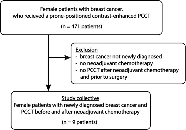
Methods: We retrospectively included patients with newly diagnosed breast cancer who received contrast-enhanced thoracic PCCT in prone position before and after NAST. Three experienced radiologists measured tumor size, tumor area, iodine uptake within tumors, number of suspicious breast lesions and of suspicious axillary lymph nodes before and after NAST. We compared the initial tumor size to contrast-enhanced magnetic resonance imaging (MRI), the residual tumor size after NAST to histopathology.
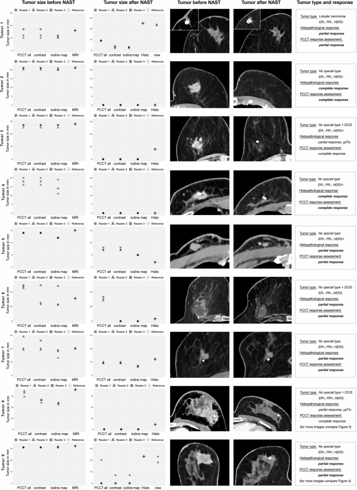
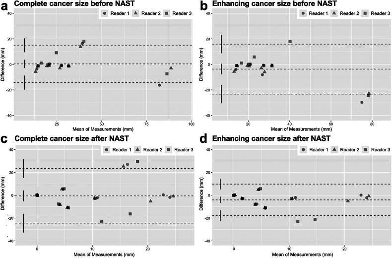
Results: Eighteen PCCT exams in nine patients aged 58 ± 14 years (mean ± standard deviation) were analyzed. After NAST, PCCT correctly identified a reduction in tumor burden in 9 of 9 cases and a complete response in 2 of 2 cases, with a significant reduction in tumor size, area, T-stage, number of suspicious breast lesions and of suspicious lymph nodes (p < 0.001 for all) as well as reduction in cutaneous infiltration (p = 0.010). Mean and maximum iodine uptake showed a nonsignificant reduction in cases with residual tumor after NAST (p = 0.092 and 0.363).
Conclusion: These preliminary findings suggest that thoracic PCCT can accurately detect local changes in breast cancer after NAST.
Relevance statement: Thoracic PCCT offers promising potential for accurately assessing breast cancer response to NAST.
Trial registration: German Clinical Trials Register DRKS00028997.
Key points: Prone thoracic contrast-enhanced photon-counting detector computed tomography (PCCT) can accurately detect reductions in tumor size, area, and T-stage. Prone PCCT can identify a decrease in the number of suspicious axillary lymph nodes. This technique shows promising results in identifying breast cancer response to neoadjuvant systemic therapy (NAST).

 求助内容:
求助内容: 应助结果提醒方式:


