{"title":"胰腺导管腺癌MRI特征与KRAS突变状态的相关性研究。","authors":"Junjian Shen, Xingxing Wang, Keqin Yu, Kai Liu, Xiaolin Wang, Haitao Sun, Jianjun Zhou, Mengsu Zeng","doi":"10.1007/s00261-025-04888-x","DOIUrl":null,"url":null,"abstract":"<div><h3>Purpose</h3><p>To investigate MRI features associated with <i>KRAS</i> mutation status in PDAC and their clinical implications.</p><h3>Materials and methods</h3><p>In our study, 1474 patients pathologically confirmed PDAC patients between January 2016 and December 2023 were evaluated. Patients with genetic testing (<i>KRAS</i> mutation status) and MRI examination were enrolled and grouped as <i>KRAS</i>-mutated PDAC and non-<i>KRAS</i>-mutated PDAC. Contrast-enhanced MRI features, clinicopathologic findings, and prognosis were compared between two groups.</p><h3>Results</h3><p>A total of 308 surgically confirmed PDAC patients (median age, 67 years [IQR, 59, 72]; 183 male and 125 female) with genetic testing data were included, of which 258 had KRAS-mutated PDAC and 50 had non-KRAS-mutated PDAC. KRAS-mutated PDAC demonstrated distinct clinicopathological characteristics, including higher rates of diabetes (OR, 2.450, 95% CI, 1.151–5.212, <i>P</i> = 0.020), pathological peripheral nerve infiltration (OR, 2.296, 95% CI, 1.083–4.867, <i>P</i> = 0.030), and pN stage (OR, 2.006, 95% CI, 1.012–3.976, <i>P</i> = 0.046). The 1-, 3-, 5-year OS rate was worse for <i>KRAS</i>-mutated PDAC (89.9%, 45.4%, 23.2% vs. 95.1%, 60.4% 60.4%, <i>P</i> = 0.045). Rim enhancement (OR = 2.039, 95% CI: 1.053, 3.951, <i>P</i> = 0.035) and larger tumor size (OR = 3.286, 95% CI: 1.523, 7.089, <i>P</i> = 0.002) were identified as distinctive MRI features for <i>KRAS</i>-mutated PDAC.</p><h3>Conclusion</h3><p><i>KRAS</i>-mutated PDAC presents unique clinical and pathological features and is associated with poorer prognosis. Rim enhancement and larger tumor size on MRI were identified as features associated with KRAS-mutated PDAC.</p><h3>Graphical abstract</h3><div><figure><div><div><picture><source><img></source></picture></div></div></figure></div></div>","PeriodicalId":7126,"journal":{"name":"Abdominal Radiology","volume":"50 10","pages":"4575 - 4588"},"PeriodicalIF":2.2000,"publicationDate":"2025-03-29","publicationTypes":"Journal Article","fieldsOfStudy":null,"isOpenAccess":false,"openAccessPdf":"","citationCount":"0","resultStr":"{\"title\":\"Correlation of MRI characteristics with KRAS mutation status in pancreatic ductal adenocarcinoma\",\"authors\":\"Junjian Shen, Xingxing Wang, Keqin Yu, Kai Liu, Xiaolin Wang, Haitao Sun, Jianjun Zhou, Mengsu Zeng\",\"doi\":\"10.1007/s00261-025-04888-x\",\"DOIUrl\":null,\"url\":null,\"abstract\":\"<div><h3>Purpose</h3><p>To investigate MRI features associated with <i>KRAS</i> mutation status in PDAC and their clinical implications.</p><h3>Materials and methods</h3><p>In our study, 1474 patients pathologically confirmed PDAC patients between January 2016 and December 2023 were evaluated. Patients with genetic testing (<i>KRAS</i> mutation status) and MRI examination were enrolled and grouped as <i>KRAS</i>-mutated PDAC and non-<i>KRAS</i>-mutated PDAC. Contrast-enhanced MRI features, clinicopathologic findings, and prognosis were compared between two groups.</p><h3>Results</h3><p>A total of 308 surgically confirmed PDAC patients (median age, 67 years [IQR, 59, 72]; 183 male and 125 female) with genetic testing data were included, of which 258 had KRAS-mutated PDAC and 50 had non-KRAS-mutated PDAC. KRAS-mutated PDAC demonstrated distinct clinicopathological characteristics, including higher rates of diabetes (OR, 2.450, 95% CI, 1.151–5.212, <i>P</i> = 0.020), pathological peripheral nerve infiltration (OR, 2.296, 95% CI, 1.083–4.867, <i>P</i> = 0.030), and pN stage (OR, 2.006, 95% CI, 1.012–3.976, <i>P</i> = 0.046). The 1-, 3-, 5-year OS rate was worse for <i>KRAS</i>-mutated PDAC (89.9%, 45.4%, 23.2% vs. 95.1%, 60.4% 60.4%, <i>P</i> = 0.045). Rim enhancement (OR = 2.039, 95% CI: 1.053, 3.951, <i>P</i> = 0.035) and larger tumor size (OR = 3.286, 95% CI: 1.523, 7.089, <i>P</i> = 0.002) were identified as distinctive MRI features for <i>KRAS</i>-mutated PDAC.</p><h3>Conclusion</h3><p><i>KRAS</i>-mutated PDAC presents unique clinical and pathological features and is associated with poorer prognosis. Rim enhancement and larger tumor size on MRI were identified as features associated with KRAS-mutated PDAC.</p><h3>Graphical abstract</h3><div><figure><div><div><picture><source><img></source></picture></div></div></figure></div></div>\",\"PeriodicalId\":7126,\"journal\":{\"name\":\"Abdominal Radiology\",\"volume\":\"50 10\",\"pages\":\"4575 - 4588\"},\"PeriodicalIF\":2.2000,\"publicationDate\":\"2025-03-29\",\"publicationTypes\":\"Journal Article\",\"fieldsOfStudy\":null,\"isOpenAccess\":false,\"openAccessPdf\":\"\",\"citationCount\":\"0\",\"resultStr\":null,\"platform\":\"Semanticscholar\",\"paperid\":null,\"PeriodicalName\":\"Abdominal Radiology\",\"FirstCategoryId\":\"3\",\"ListUrlMain\":\"https://link.springer.com/article/10.1007/s00261-025-04888-x\",\"RegionNum\":3,\"RegionCategory\":\"医学\",\"ArticlePicture\":[],\"TitleCN\":null,\"AbstractTextCN\":null,\"PMCID\":null,\"EPubDate\":\"\",\"PubModel\":\"\",\"JCR\":\"Q2\",\"JCRName\":\"RADIOLOGY, NUCLEAR MEDICINE & MEDICAL IMAGING\",\"Score\":null,\"Total\":0}","platform":"Semanticscholar","paperid":null,"PeriodicalName":"Abdominal Radiology","FirstCategoryId":"3","ListUrlMain":"https://link.springer.com/article/10.1007/s00261-025-04888-x","RegionNum":3,"RegionCategory":"医学","ArticlePicture":[],"TitleCN":null,"AbstractTextCN":null,"PMCID":null,"EPubDate":"","PubModel":"","JCR":"Q2","JCRName":"RADIOLOGY, NUCLEAR MEDICINE & MEDICAL IMAGING","Score":null,"Total":0}
引用次数: 0
摘要
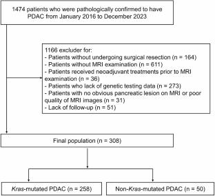
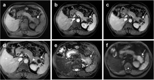
目的:探讨与PDAC中KRAS突变状态相关的MRI特征及其临床意义。材料与方法:本研究对2016年1月至2023年12月1474例病理证实的PDAC患者进行评估。纳入基因检测(KRAS突变状态)和MRI检查的患者,并将其分为KRAS突变PDAC和非KRAS突变PDAC。比较两组患者的MRI增强特征、临床病理表现及预后。结果:308例经手术确诊的PDAC患者(中位年龄67岁[IQR, 59, 72];纳入183例(男125例,女125例)基因检测资料,其中258例为kras突变PDAC, 50例为非kras突变PDAC。kras突变的PDAC表现出明显的临床病理特征,包括较高的糖尿病发生率(OR, 2.450, 95% CI, 1.151-5.212, P = 0.020)、病理性周围神经浸润(OR, 2.296, 95% CI, 1.083-4.867, P = 0.030)和pN分期(OR, 2.006, 95% CI, 1.012-3.976, P = 0.046)。kras突变PDAC的1、3、5年OS率较差(89.9%、45.4%、23.2% vs. 95.1%、60.4%、60.4%,P = 0.045)。环增强(OR = 2.039, 95% CI: 1.053, 3.951, P = 0.035)和较大的肿瘤大小(OR = 3.286, 95% CI: 1.523, 7.089, P = 0.002)被确定为kras突变PDAC的独特MRI特征。结论:kras突变的PDAC具有独特的临床和病理特征,与较差的预后相关。MRI上的边缘增强和更大的肿瘤大小被确定为与kras突变的PDAC相关的特征。
Correlation of MRI characteristics with KRAS mutation status in pancreatic ductal adenocarcinoma
Purpose
To investigate MRI features associated with KRAS mutation status in PDAC and their clinical implications.
Materials and methods
In our study, 1474 patients pathologically confirmed PDAC patients between January 2016 and December 2023 were evaluated. Patients with genetic testing (KRAS mutation status) and MRI examination were enrolled and grouped as KRAS-mutated PDAC and non-KRAS-mutated PDAC. Contrast-enhanced MRI features, clinicopathologic findings, and prognosis were compared between two groups.
Results
A total of 308 surgically confirmed PDAC patients (median age, 67 years [IQR, 59, 72]; 183 male and 125 female) with genetic testing data were included, of which 258 had KRAS-mutated PDAC and 50 had non-KRAS-mutated PDAC. KRAS-mutated PDAC demonstrated distinct clinicopathological characteristics, including higher rates of diabetes (OR, 2.450, 95% CI, 1.151–5.212, P = 0.020), pathological peripheral nerve infiltration (OR, 2.296, 95% CI, 1.083–4.867, P = 0.030), and pN stage (OR, 2.006, 95% CI, 1.012–3.976, P = 0.046). The 1-, 3-, 5-year OS rate was worse for KRAS-mutated PDAC (89.9%, 45.4%, 23.2% vs. 95.1%, 60.4% 60.4%, P = 0.045). Rim enhancement (OR = 2.039, 95% CI: 1.053, 3.951, P = 0.035) and larger tumor size (OR = 3.286, 95% CI: 1.523, 7.089, P = 0.002) were identified as distinctive MRI features for KRAS-mutated PDAC.
Conclusion
KRAS-mutated PDAC presents unique clinical and pathological features and is associated with poorer prognosis. Rim enhancement and larger tumor size on MRI were identified as features associated with KRAS-mutated PDAC.
期刊介绍:
Abdominal Radiology seeks to meet the professional needs of the abdominal radiologist by publishing clinically pertinent original, review and practice related articles on the gastrointestinal and genitourinary tracts and abdominal interventional and radiologic procedures. Case reports are generally not accepted unless they are the first report of a new disease or condition, or part of a special solicited section.
Reasons to Publish Your Article in Abdominal Radiology:
· Official journal of the Society of Abdominal Radiology (SAR)
· Published in Cooperation with:
European Society of Gastrointestinal and Abdominal Radiology (ESGAR)
European Society of Urogenital Radiology (ESUR)
Asian Society of Abdominal Radiology (ASAR)
· Efficient handling and Expeditious review
· Author feedback is provided in a mentoring style
· Global readership
· Readers can earn CME credits

 求助内容:
求助内容: 应助结果提醒方式:


