Matthias Maak, Philipp Mörsdorf, Layla Bari, Myriam Braun-Münker, Maximilian Scharonow, Marcel Orth, Dietrich Doll
{"title":"臀间沟深度对毛突窦疾病的发展无影响。","authors":"Matthias Maak, Philipp Mörsdorf, Layla Bari, Myriam Braun-Münker, Maximilian Scharonow, Marcel Orth, Dietrich Doll","doi":"10.47717/turkjsurg.2025.6665","DOIUrl":null,"url":null,"abstract":"<p><strong>Objective: </strong>The etiology of primary pilonidal sinus disease (PSD) remains unclear. Prior investigations suggest that sharp fragments from the occiput contribute to the formation of PSD. In 2009 a correlation between PSD and a deeper natal cleft was reported. We investigated the association between intergluteal fold (IGF) depth and PSD risk using a standardized five-step measuring protocol.</p><p><strong>Material and methods: </strong>Our clinical prospective study included 95 PSD patients and 105 non-PSD individuals, and measurements were taken from the glabella sacralis to the anus in a northern German population.</p><p><strong>Results: </strong>The mean (± standard deviation) intergluteal depth progressively increased from the intergluteal opening from the sacral glabella at 9.1 (±3.4) mm to a maximum of 62.6 (±10.4) mm. Notably, the deepest point was consistently observed at the anus, where PSD occurrence is rare. No significant difference in IGF depth between PSD and non-PSD patients was found. Additionally, PSD predominantly developed in the proximal (cranial) third of the IGF, despite the maximum depth being in the distal region.</p><p><strong>Conclusion: </strong>These findings suggest that IGF depth is not a risk factor for PSD.</p>","PeriodicalId":23374,"journal":{"name":"Turkish Journal of Surgery","volume":" ","pages":"130-134"},"PeriodicalIF":0.6000,"publicationDate":"2025-05-30","publicationTypes":"Journal Article","fieldsOfStudy":null,"isOpenAccess":false,"openAccessPdf":"https://www.ncbi.nlm.nih.gov/pmc/articles/PMC12124332/pdf/","citationCount":"0","resultStr":"{\"title\":\"Intergluteal fold depth has no influence on pilonidal sinus disease development.\",\"authors\":\"Matthias Maak, Philipp Mörsdorf, Layla Bari, Myriam Braun-Münker, Maximilian Scharonow, Marcel Orth, Dietrich Doll\",\"doi\":\"10.47717/turkjsurg.2025.6665\",\"DOIUrl\":null,\"url\":null,\"abstract\":\"<p><strong>Objective: </strong>The etiology of primary pilonidal sinus disease (PSD) remains unclear. Prior investigations suggest that sharp fragments from the occiput contribute to the formation of PSD. In 2009 a correlation between PSD and a deeper natal cleft was reported. We investigated the association between intergluteal fold (IGF) depth and PSD risk using a standardized five-step measuring protocol.</p><p><strong>Material and methods: </strong>Our clinical prospective study included 95 PSD patients and 105 non-PSD individuals, and measurements were taken from the glabella sacralis to the anus in a northern German population.</p><p><strong>Results: </strong>The mean (± standard deviation) intergluteal depth progressively increased from the intergluteal opening from the sacral glabella at 9.1 (±3.4) mm to a maximum of 62.6 (±10.4) mm. Notably, the deepest point was consistently observed at the anus, where PSD occurrence is rare. No significant difference in IGF depth between PSD and non-PSD patients was found. Additionally, PSD predominantly developed in the proximal (cranial) third of the IGF, despite the maximum depth being in the distal region.</p><p><strong>Conclusion: </strong>These findings suggest that IGF depth is not a risk factor for PSD.</p>\",\"PeriodicalId\":23374,\"journal\":{\"name\":\"Turkish Journal of Surgery\",\"volume\":\" \",\"pages\":\"130-134\"},\"PeriodicalIF\":0.6000,\"publicationDate\":\"2025-05-30\",\"publicationTypes\":\"Journal Article\",\"fieldsOfStudy\":null,\"isOpenAccess\":false,\"openAccessPdf\":\"https://www.ncbi.nlm.nih.gov/pmc/articles/PMC12124332/pdf/\",\"citationCount\":\"0\",\"resultStr\":null,\"platform\":\"Semanticscholar\",\"paperid\":null,\"PeriodicalName\":\"Turkish Journal of Surgery\",\"FirstCategoryId\":\"1085\",\"ListUrlMain\":\"https://doi.org/10.47717/turkjsurg.2025.6665\",\"RegionNum\":0,\"RegionCategory\":null,\"ArticlePicture\":[],\"TitleCN\":null,\"AbstractTextCN\":null,\"PMCID\":null,\"EPubDate\":\"2025/3/28 0:00:00\",\"PubModel\":\"Epub\",\"JCR\":\"Q4\",\"JCRName\":\"SURGERY\",\"Score\":null,\"Total\":0}","platform":"Semanticscholar","paperid":null,"PeriodicalName":"Turkish Journal of Surgery","FirstCategoryId":"1085","ListUrlMain":"https://doi.org/10.47717/turkjsurg.2025.6665","RegionNum":0,"RegionCategory":null,"ArticlePicture":[],"TitleCN":null,"AbstractTextCN":null,"PMCID":null,"EPubDate":"2025/3/28 0:00:00","PubModel":"Epub","JCR":"Q4","JCRName":"SURGERY","Score":null,"Total":0}
Intergluteal fold depth has no influence on pilonidal sinus disease development.
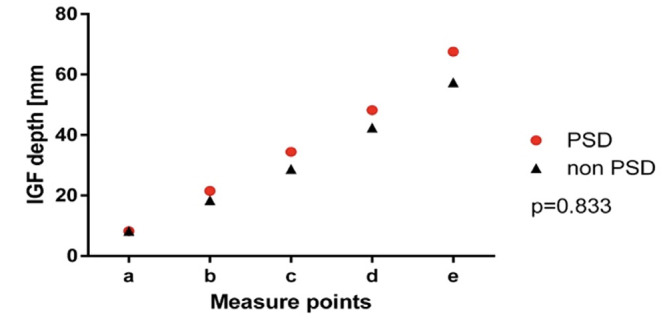
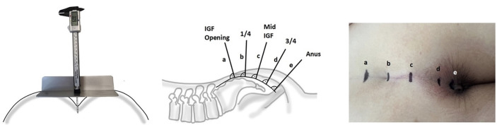
Objective: The etiology of primary pilonidal sinus disease (PSD) remains unclear. Prior investigations suggest that sharp fragments from the occiput contribute to the formation of PSD. In 2009 a correlation between PSD and a deeper natal cleft was reported. We investigated the association between intergluteal fold (IGF) depth and PSD risk using a standardized five-step measuring protocol.
Material and methods: Our clinical prospective study included 95 PSD patients and 105 non-PSD individuals, and measurements were taken from the glabella sacralis to the anus in a northern German population.
Results: The mean (± standard deviation) intergluteal depth progressively increased from the intergluteal opening from the sacral glabella at 9.1 (±3.4) mm to a maximum of 62.6 (±10.4) mm. Notably, the deepest point was consistently observed at the anus, where PSD occurrence is rare. No significant difference in IGF depth between PSD and non-PSD patients was found. Additionally, PSD predominantly developed in the proximal (cranial) third of the IGF, despite the maximum depth being in the distal region.
Conclusion: These findings suggest that IGF depth is not a risk factor for PSD.

 求助内容:
求助内容: 应助结果提醒方式:


