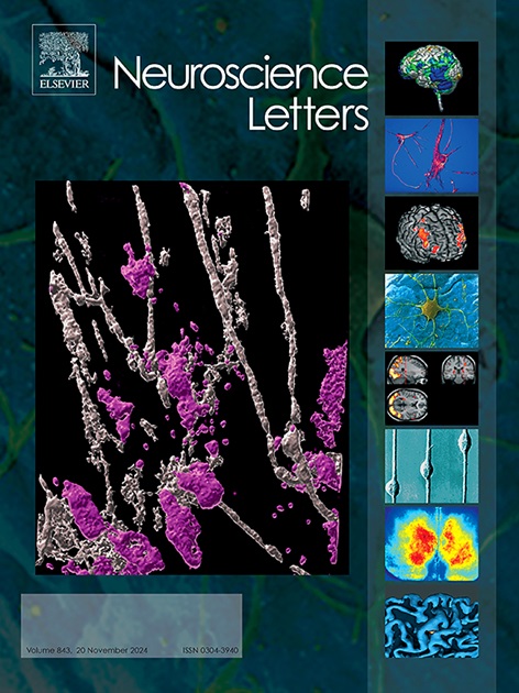
阿尔茨海默病大鼠海马内观察到的金胶体聚集体的低聚物敏感原位检测和表征。
IF 2.5
4区 医学
Q3 NEUROSCIENCES
引用次数: 0
摘要
为了更好地了解大鼠模型中导致阿尔茨海默病(AD)的病理低聚物的形成动态,本研究对海马中金胶体上聚集的蛋白质进行了检测。Long Evans Cohen's AD(+)大鼠模型的海马切片与金胶体混合,并通过表面增强拉曼散射(SERS)成像对所产生的聚集体进行了检测。与AD(-)大鼠组织相比,AD(+)大鼠海马组织产生的金胶体聚集体更大。每个海马切片的SERS光谱在酰胺I、II和III波段区域显示出相似的光谱模式,但在AD(+)大鼠组织的300 cm-1 - 1250 cm-1区域分别显示出不同的光谱模式。此前曾有报道称,具有β片构象的淀粉样纤维会在小鼠和人类AD脑组织中形成金胶体聚集体。与观察到的 AD(-)大鼠相比,AD(+)大鼠海马脑切片中的金胶体聚集体表现出明显的形态特征。这表明低聚物浓度在海马中存在空间分布,从而诱导纤维形成,破坏海马内部和大脑其他部分之间的神经元网络。本文章由计算机程序翻译,如有差异,请以英文原文为准。
Oligomer sensitive in-situ detection and characterization of gold colloid aggregate formations observed within the hippocampus of the Alzheimer’s disease rat
In order to better understand the dynamics governing the formation of pathological oligomers leading to Alzheimer’s disease (AD) in a rat model the present study examined the protein aggregates accumulating on gold colloids in the hippocampus. Sections of the hippocampus of the Long Evans Cohen’s AD(+) rat model were mixed with gold colloids and the resulting aggregates were examined by Surface Enhanced Raman Scattering (SERS) imaging. Compared to AD(–) rat tissues, the AD(+) rat hippocampal tissues produced a larger sized gold colloid aggregates. The SERS spectrum of each hippocampal section exhibited similar spectral patterns in the Amide I, II, and III band regions, but showed distinct spectral patterns in the region between 300 cm−1 – 1250 cm−1 in AD(+) rat tissues, respectively. Amyloid fibrils with a β-sheet conformation were previously reported to form gold colloid aggregates in mouse and human AD brain tissues. The gold colloid aggregates in the AD (+) rat hippocampal brain sections showed distinct morphological traits compared to those observed in AD(–) rats. This suggests that there is a spatial distribution of oligomer concentration in the hippocampus, which induces fibril formation to disrupt neuronal networks within the hippocampus and between other parts of the brain.
求助全文
通过发布文献求助,成功后即可免费获取论文全文。
去求助
来源期刊

Neuroscience Letters
医学-神经科学
CiteScore
5.20
自引率
0.00%
发文量
408
审稿时长
50 days
期刊介绍:
Neuroscience Letters is devoted to the rapid publication of short, high-quality papers of interest to the broad community of neuroscientists. Only papers which will make a significant addition to the literature in the field will be published. Papers in all areas of neuroscience - molecular, cellular, developmental, systems, behavioral and cognitive, as well as computational - will be considered for publication. Submission of laboratory investigations that shed light on disease mechanisms is encouraged. Special Issues, edited by Guest Editors to cover new and rapidly-moving areas, will include invited mini-reviews. Occasional mini-reviews in especially timely areas will be considered for publication, without invitation, outside of Special Issues; these un-solicited mini-reviews can be submitted without invitation but must be of very high quality. Clinical studies will also be published if they provide new information about organization or actions of the nervous system, or provide new insights into the neurobiology of disease. NSL does not publish case reports.
 求助内容:
求助内容: 应助结果提醒方式:


