Daniel I. Glazer, Melissa Viator, Andrew Sharp, Jay B. Patel, Borna E. Dabiri, Christopher P. Bridge, Justine A. Barletta, Oleg S. Pianykh, William W. Mayo-Smith
{"title":"利用可解释的规则学习人工智能与CT放射组学最佳区分肾上腺嗜铬细胞瘤和腺瘤。","authors":"Daniel I. Glazer, Melissa Viator, Andrew Sharp, Jay B. Patel, Borna E. Dabiri, Christopher P. Bridge, Justine A. Barletta, Oleg S. Pianykh, William W. Mayo-Smith","doi":"10.1007/s00261-025-04893-0","DOIUrl":null,"url":null,"abstract":"<div><h3>Purpose</h3><p>To identify interpretable CT-based radiomics features that can differentiate adrenal pheochromocytomas from adenomas.</p><h3>Methods</h3><p>An institutional database was used to identify patients with pathologically proven adrenal pheochromocytomas 5/1/05–5/1/23. To be included, patients needed to have a contrast-enhanced abdominal CT with an adrenal mass within 12 months of pathology (<i>n</i> = 95). For comparison, 57 adenomas were identified from a set of consecutive CT examinations. The final dataset included 152 adrenal masses (95 pheochromocytomas; 57 adenomas) with 121 used in the development set and 31 in the test set. Following confirmation of accurate automated segmentation, 463 radiomic features were evaluated and used to create an interpretable artificial intelligence (AI) rule-learning model. Model performance was reported using F1 score.</p><h3>Results</h3><p>The study included 146 patients (age 59 years +/- 21; 89 females). A three-feature rule, High Gray Level Zone Emphasis > 184, Roundness > 0.35, and Boundary Low Gray Level Emphasis < 0.021 produced an F1 score of 0.97 on the train set (95% confidence interval [CI]: 0.94, 0.99) and 0.96 on the test set (95% CI: 0.89, 1.00). The rule-learning model determined that the rule most predictive of pheochromocytoma was Maximum Pixel Attenuation > 125 HU resulting in an F1 score of 0.89 (95% CI: 0.83, 0.94) on the training set and 0.93 (95% CI: 0.83, 0.99) on the test set.</p><h3>Conclusion</h3><p>A rule-learning AI model identified the smallest optimal set of interpretable CT radiomics features, sufficient to achieve 96% accuracy in differentiating adrenal pheochromocytomas from adenomas on contrast enhanced CT.</p><h3>Graphical abstract</h3><div><figure><div><div><picture><source><img></source></picture></div></div></figure></div></div>","PeriodicalId":7126,"journal":{"name":"Abdominal Radiology","volume":"50 10","pages":"4722 - 4730"},"PeriodicalIF":2.2000,"publicationDate":"2025-03-26","publicationTypes":"Journal Article","fieldsOfStudy":null,"isOpenAccess":false,"openAccessPdf":"","citationCount":"0","resultStr":"{\"title\":\"Using interpretable rule-learning artificial intelligence to optimally differentiate adrenal pheochromocytomas from adenomas with CT radiomics\",\"authors\":\"Daniel I. Glazer, Melissa Viator, Andrew Sharp, Jay B. Patel, Borna E. Dabiri, Christopher P. Bridge, Justine A. Barletta, Oleg S. Pianykh, William W. Mayo-Smith\",\"doi\":\"10.1007/s00261-025-04893-0\",\"DOIUrl\":null,\"url\":null,\"abstract\":\"<div><h3>Purpose</h3><p>To identify interpretable CT-based radiomics features that can differentiate adrenal pheochromocytomas from adenomas.</p><h3>Methods</h3><p>An institutional database was used to identify patients with pathologically proven adrenal pheochromocytomas 5/1/05–5/1/23. To be included, patients needed to have a contrast-enhanced abdominal CT with an adrenal mass within 12 months of pathology (<i>n</i> = 95). For comparison, 57 adenomas were identified from a set of consecutive CT examinations. The final dataset included 152 adrenal masses (95 pheochromocytomas; 57 adenomas) with 121 used in the development set and 31 in the test set. Following confirmation of accurate automated segmentation, 463 radiomic features were evaluated and used to create an interpretable artificial intelligence (AI) rule-learning model. Model performance was reported using F1 score.</p><h3>Results</h3><p>The study included 146 patients (age 59 years +/- 21; 89 females). A three-feature rule, High Gray Level Zone Emphasis > 184, Roundness > 0.35, and Boundary Low Gray Level Emphasis < 0.021 produced an F1 score of 0.97 on the train set (95% confidence interval [CI]: 0.94, 0.99) and 0.96 on the test set (95% CI: 0.89, 1.00). The rule-learning model determined that the rule most predictive of pheochromocytoma was Maximum Pixel Attenuation > 125 HU resulting in an F1 score of 0.89 (95% CI: 0.83, 0.94) on the training set and 0.93 (95% CI: 0.83, 0.99) on the test set.</p><h3>Conclusion</h3><p>A rule-learning AI model identified the smallest optimal set of interpretable CT radiomics features, sufficient to achieve 96% accuracy in differentiating adrenal pheochromocytomas from adenomas on contrast enhanced CT.</p><h3>Graphical abstract</h3><div><figure><div><div><picture><source><img></source></picture></div></div></figure></div></div>\",\"PeriodicalId\":7126,\"journal\":{\"name\":\"Abdominal Radiology\",\"volume\":\"50 10\",\"pages\":\"4722 - 4730\"},\"PeriodicalIF\":2.2000,\"publicationDate\":\"2025-03-26\",\"publicationTypes\":\"Journal Article\",\"fieldsOfStudy\":null,\"isOpenAccess\":false,\"openAccessPdf\":\"\",\"citationCount\":\"0\",\"resultStr\":null,\"platform\":\"Semanticscholar\",\"paperid\":null,\"PeriodicalName\":\"Abdominal Radiology\",\"FirstCategoryId\":\"3\",\"ListUrlMain\":\"https://link.springer.com/article/10.1007/s00261-025-04893-0\",\"RegionNum\":3,\"RegionCategory\":\"医学\",\"ArticlePicture\":[],\"TitleCN\":null,\"AbstractTextCN\":null,\"PMCID\":null,\"EPubDate\":\"\",\"PubModel\":\"\",\"JCR\":\"Q2\",\"JCRName\":\"RADIOLOGY, NUCLEAR MEDICINE & MEDICAL IMAGING\",\"Score\":null,\"Total\":0}","platform":"Semanticscholar","paperid":null,"PeriodicalName":"Abdominal Radiology","FirstCategoryId":"3","ListUrlMain":"https://link.springer.com/article/10.1007/s00261-025-04893-0","RegionNum":3,"RegionCategory":"医学","ArticlePicture":[],"TitleCN":null,"AbstractTextCN":null,"PMCID":null,"EPubDate":"","PubModel":"","JCR":"Q2","JCRName":"RADIOLOGY, NUCLEAR MEDICINE & MEDICAL IMAGING","Score":null,"Total":0}
Using interpretable rule-learning artificial intelligence to optimally differentiate adrenal pheochromocytomas from adenomas with CT radiomics
Purpose
To identify interpretable CT-based radiomics features that can differentiate adrenal pheochromocytomas from adenomas.
Methods
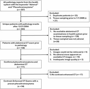
An institutional database was used to identify patients with pathologically proven adrenal pheochromocytomas 5/1/05–5/1/23. To be included, patients needed to have a contrast-enhanced abdominal CT with an adrenal mass within 12 months of pathology (n = 95). For comparison, 57 adenomas were identified from a set of consecutive CT examinations. The final dataset included 152 adrenal masses (95 pheochromocytomas; 57 adenomas) with 121 used in the development set and 31 in the test set. Following confirmation of accurate automated segmentation, 463 radiomic features were evaluated and used to create an interpretable artificial intelligence (AI) rule-learning model. Model performance was reported using F1 score.
Results
The study included 146 patients (age 59 years +/- 21; 89 females). A three-feature rule, High Gray Level Zone Emphasis > 184, Roundness > 0.35, and Boundary Low Gray Level Emphasis < 0.021 produced an F1 score of 0.97 on the train set (95% confidence interval [CI]: 0.94, 0.99) and 0.96 on the test set (95% CI: 0.89, 1.00). The rule-learning model determined that the rule most predictive of pheochromocytoma was Maximum Pixel Attenuation > 125 HU resulting in an F1 score of 0.89 (95% CI: 0.83, 0.94) on the training set and 0.93 (95% CI: 0.83, 0.99) on the test set.
Conclusion
A rule-learning AI model identified the smallest optimal set of interpretable CT radiomics features, sufficient to achieve 96% accuracy in differentiating adrenal pheochromocytomas from adenomas on contrast enhanced CT.
期刊介绍:
Abdominal Radiology seeks to meet the professional needs of the abdominal radiologist by publishing clinically pertinent original, review and practice related articles on the gastrointestinal and genitourinary tracts and abdominal interventional and radiologic procedures. Case reports are generally not accepted unless they are the first report of a new disease or condition, or part of a special solicited section.
Reasons to Publish Your Article in Abdominal Radiology:
· Official journal of the Society of Abdominal Radiology (SAR)
· Published in Cooperation with:
European Society of Gastrointestinal and Abdominal Radiology (ESGAR)
European Society of Urogenital Radiology (ESUR)
Asian Society of Abdominal Radiology (ASAR)
· Efficient handling and Expeditious review
· Author feedback is provided in a mentoring style
· Global readership
· Readers can earn CME credits


 求助内容:
求助内容: 应助结果提醒方式:


