Zachary Corey, Lydia A Luu, Sabrina Newman, Shyam S Raghavan
{"title":"心脏淀粉样变性心脏移植后免疫抑制患者左前臂紫色结节。","authors":"Zachary Corey, Lydia A Luu, Sabrina Newman, Shyam S Raghavan","doi":"10.3390/dermatopathology12010002","DOIUrl":null,"url":null,"abstract":"<p><p>We present the case of a 60-year-old immunocompromised man who presented with two pruritic pink-red indurated nodules with overlying scale and focal areas of ulceration on his left dorsal and left medial forearm, which evolved over a 2-month period. The pathology showed numerous fungal hyphae present that were pauci-septate with various branched angles and variable hyphal thickness. Fungal cultures grew <i>Rhizopus</i> species and a universal fungal PCR detected the <i>Rhizopus oryzae</i> complex. Based on the clinicopathologic correlation, the diagnosis of cutaneous mucormycosis was made. Cutaneous mucormycosis is an aggressive fungal infection of the <i>Mucorales</i> family occurring after the inoculation of fungal spores in disrupted skin. It usually presents as a necrotic eschar but can also present as cellulitis that evolves into a necrotic ulcer. A prompt diagnosis is critical for the effective management of cutaneous mucormycosis. The treatment includes an immediate systemic treatment with amphotericin B and a surgical debridement of the necrotic regions. Given the wide range of presenting symptoms, clinical suspicion for this emergent condition must remain high in immunocompromised and diabetic patients.</p>","PeriodicalId":42885,"journal":{"name":"Dermatopathology","volume":"12 1","pages":""},"PeriodicalIF":1.7000,"publicationDate":"2025-01-16","publicationTypes":"Journal Article","fieldsOfStudy":null,"isOpenAccess":false,"openAccessPdf":"https://www.ncbi.nlm.nih.gov/pmc/articles/PMC11755463/pdf/","citationCount":"0","resultStr":"{\"title\":\"Violaceous Nodules on the Left Forearm of an Immunosuppressed Patient Following Heart Transplantation for Cardiac Amyloidosis.\",\"authors\":\"Zachary Corey, Lydia A Luu, Sabrina Newman, Shyam S Raghavan\",\"doi\":\"10.3390/dermatopathology12010002\",\"DOIUrl\":null,\"url\":null,\"abstract\":\"<p><p>We present the case of a 60-year-old immunocompromised man who presented with two pruritic pink-red indurated nodules with overlying scale and focal areas of ulceration on his left dorsal and left medial forearm, which evolved over a 2-month period. The pathology showed numerous fungal hyphae present that were pauci-septate with various branched angles and variable hyphal thickness. Fungal cultures grew <i>Rhizopus</i> species and a universal fungal PCR detected the <i>Rhizopus oryzae</i> complex. Based on the clinicopathologic correlation, the diagnosis of cutaneous mucormycosis was made. Cutaneous mucormycosis is an aggressive fungal infection of the <i>Mucorales</i> family occurring after the inoculation of fungal spores in disrupted skin. It usually presents as a necrotic eschar but can also present as cellulitis that evolves into a necrotic ulcer. A prompt diagnosis is critical for the effective management of cutaneous mucormycosis. The treatment includes an immediate systemic treatment with amphotericin B and a surgical debridement of the necrotic regions. Given the wide range of presenting symptoms, clinical suspicion for this emergent condition must remain high in immunocompromised and diabetic patients.</p>\",\"PeriodicalId\":42885,\"journal\":{\"name\":\"Dermatopathology\",\"volume\":\"12 1\",\"pages\":\"\"},\"PeriodicalIF\":1.7000,\"publicationDate\":\"2025-01-16\",\"publicationTypes\":\"Journal Article\",\"fieldsOfStudy\":null,\"isOpenAccess\":false,\"openAccessPdf\":\"https://www.ncbi.nlm.nih.gov/pmc/articles/PMC11755463/pdf/\",\"citationCount\":\"0\",\"resultStr\":null,\"platform\":\"Semanticscholar\",\"paperid\":null,\"PeriodicalName\":\"Dermatopathology\",\"FirstCategoryId\":\"1085\",\"ListUrlMain\":\"https://doi.org/10.3390/dermatopathology12010002\",\"RegionNum\":0,\"RegionCategory\":null,\"ArticlePicture\":[],\"TitleCN\":null,\"AbstractTextCN\":null,\"PMCID\":null,\"EPubDate\":\"\",\"PubModel\":\"\",\"JCR\":\"Q3\",\"JCRName\":\"DERMATOLOGY\",\"Score\":null,\"Total\":0}","platform":"Semanticscholar","paperid":null,"PeriodicalName":"Dermatopathology","FirstCategoryId":"1085","ListUrlMain":"https://doi.org/10.3390/dermatopathology12010002","RegionNum":0,"RegionCategory":null,"ArticlePicture":[],"TitleCN":null,"AbstractTextCN":null,"PMCID":null,"EPubDate":"","PubModel":"","JCR":"Q3","JCRName":"DERMATOLOGY","Score":null,"Total":0}
Violaceous Nodules on the Left Forearm of an Immunosuppressed Patient Following Heart Transplantation for Cardiac Amyloidosis.
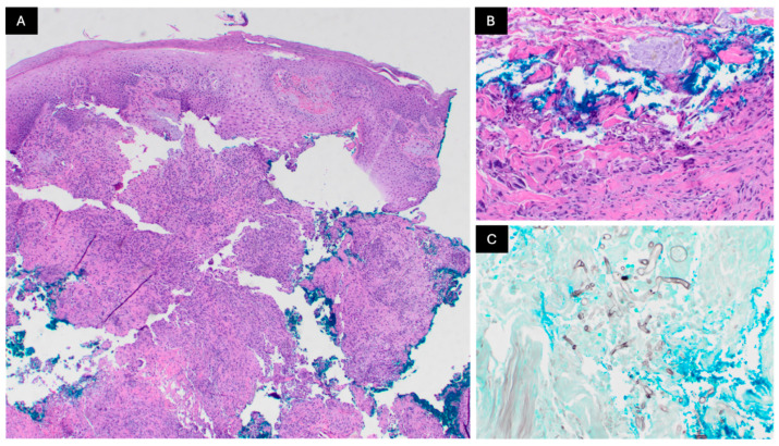
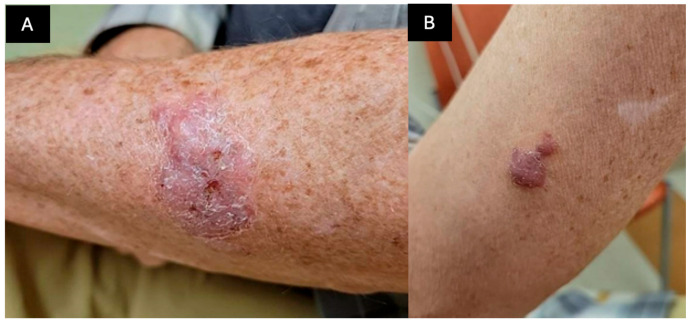
We present the case of a 60-year-old immunocompromised man who presented with two pruritic pink-red indurated nodules with overlying scale and focal areas of ulceration on his left dorsal and left medial forearm, which evolved over a 2-month period. The pathology showed numerous fungal hyphae present that were pauci-septate with various branched angles and variable hyphal thickness. Fungal cultures grew Rhizopus species and a universal fungal PCR detected the Rhizopus oryzae complex. Based on the clinicopathologic correlation, the diagnosis of cutaneous mucormycosis was made. Cutaneous mucormycosis is an aggressive fungal infection of the Mucorales family occurring after the inoculation of fungal spores in disrupted skin. It usually presents as a necrotic eschar but can also present as cellulitis that evolves into a necrotic ulcer. A prompt diagnosis is critical for the effective management of cutaneous mucormycosis. The treatment includes an immediate systemic treatment with amphotericin B and a surgical debridement of the necrotic regions. Given the wide range of presenting symptoms, clinical suspicion for this emergent condition must remain high in immunocompromised and diabetic patients.

 求助内容:
求助内容: 应助结果提醒方式:


