Luise Brock, Hadas Ben-Atya, Ashish Tiwari, Dareen Saab, Narmeen Haj, Lukas Folle, Galit Saar, Andreas Maier, Moti Freiman, Katrien Vandoorne
{"title":"改进的MRI检测炎症引起的小鼠骨髓微结构变化:机器学习增强的T2分布分析。","authors":"Luise Brock, Hadas Ben-Atya, Ashish Tiwari, Dareen Saab, Narmeen Haj, Lukas Folle, Galit Saar, Andreas Maier, Moti Freiman, Katrien Vandoorne","doi":"10.1186/s41747-025-00574-1","DOIUrl":null,"url":null,"abstract":"<p><strong>Background: </strong>We investigated inflammation-induced changes in femoral hematopoietic bone marrow using advanced magnetic resonance imaging (MRI) techniques, including T2-weighted imaging, scalar T2 mapping, and machine learning-enhanced T2 distribution analysis to improve the detection of bone marrow microstructural alterations. Findings were correlated with histological markers and systemic inflammation.</p><p><strong>Methods: </strong>Using a 9.4-T magnet, T2-weighted and multislice multiecho sequences were applied to evaluate bone marrow in female C57BL/6J mice divided into three groups: (1) controls; (2) lipopolysaccharide-induced acute inflammation (LPS); and (3) streptozotocin (STZ)- and LPS-induced diabetic inflammation (STZ + LPS). T2 relaxation times and their distributions with scalar mapping and model-informed machine learning (MIML) were analyzed. Correlations with histological iron levels and blood neutrophil counts were assessed.</p><p><strong>Results: </strong>T2-weighted imaging showed a reduced signal-to-noise ratio in inflamed bone marrow (p = 0.034). Scalar T2 mapping identified decreased T2 relaxation times (p = 0.042), moderately correlating with neutrophil counts (ρ = 0.027) and iron levels (ρ = 0.016). MIML-enhanced T2 distribution analysis exhibited superior sensitivity than scalar T2 mapping, revealing significant reductions in the first T2 distribution peak (p = 0.0025), which strongly correlated with neutrophil counts (ρ = 0.0016) and iron sequestration (ρ = 0.0002). Histology confirmed elevated iron deposits in inflamed marrow, aligning with systemic inflammation.</p><p><strong>Conclusion: </strong>Combining T2-weighted imaging, scalar T2 mapping, and MIML-enhanced T2 distribution analysis offers complementary insights into inflammation-induced bone marrow remodeling. T2 distribution analysis emerged as a more sensitive tool for detecting microstructural changes, such as iron sequestration, supporting its potential as a noninvasive biomarker for diagnosing and monitoring inflammatory diseases.</p><p><strong>Relevance statement: </strong>This study highlights the potential of advanced MRI T2 analysis and machine learning methods for noninvasive detection of inflammation-induced microstructural changes in bone marrow, offering promising diagnostic tools for inflammatory diseases.</p><p><strong>Key points: </strong>This study investigated inflammation-induced changes in bone marrow with T2 MRI and MIML. MIML outperformed quantitative scalar T2 analysis, increasingly detecting inflammation and iron sequestration in the hematopoietic bone marrow. T2 MRI with MIML analysis could aid in the early diagnosis and management of inflammatory diseases.</p>","PeriodicalId":36926,"journal":{"name":"European Radiology Experimental","volume":"9 1","pages":"39"},"PeriodicalIF":3.6000,"publicationDate":"2025-03-26","publicationTypes":"Journal Article","fieldsOfStudy":null,"isOpenAccess":false,"openAccessPdf":"https://www.ncbi.nlm.nih.gov/pmc/articles/PMC11947359/pdf/","citationCount":"0","resultStr":"{\"title\":\"Improved MRI detection of inflammation-induced changes in bone marrow microstructure in mice: a machine learning-enhanced T2 distribution analysis.\",\"authors\":\"Luise Brock, Hadas Ben-Atya, Ashish Tiwari, Dareen Saab, Narmeen Haj, Lukas Folle, Galit Saar, Andreas Maier, Moti Freiman, Katrien Vandoorne\",\"doi\":\"10.1186/s41747-025-00574-1\",\"DOIUrl\":null,\"url\":null,\"abstract\":\"<p><strong>Background: </strong>We investigated inflammation-induced changes in femoral hematopoietic bone marrow using advanced magnetic resonance imaging (MRI) techniques, including T2-weighted imaging, scalar T2 mapping, and machine learning-enhanced T2 distribution analysis to improve the detection of bone marrow microstructural alterations. Findings were correlated with histological markers and systemic inflammation.</p><p><strong>Methods: </strong>Using a 9.4-T magnet, T2-weighted and multislice multiecho sequences were applied to evaluate bone marrow in female C57BL/6J mice divided into three groups: (1) controls; (2) lipopolysaccharide-induced acute inflammation (LPS); and (3) streptozotocin (STZ)- and LPS-induced diabetic inflammation (STZ + LPS). T2 relaxation times and their distributions with scalar mapping and model-informed machine learning (MIML) were analyzed. Correlations with histological iron levels and blood neutrophil counts were assessed.</p><p><strong>Results: </strong>T2-weighted imaging showed a reduced signal-to-noise ratio in inflamed bone marrow (p = 0.034). Scalar T2 mapping identified decreased T2 relaxation times (p = 0.042), moderately correlating with neutrophil counts (ρ = 0.027) and iron levels (ρ = 0.016). MIML-enhanced T2 distribution analysis exhibited superior sensitivity than scalar T2 mapping, revealing significant reductions in the first T2 distribution peak (p = 0.0025), which strongly correlated with neutrophil counts (ρ = 0.0016) and iron sequestration (ρ = 0.0002). Histology confirmed elevated iron deposits in inflamed marrow, aligning with systemic inflammation.</p><p><strong>Conclusion: </strong>Combining T2-weighted imaging, scalar T2 mapping, and MIML-enhanced T2 distribution analysis offers complementary insights into inflammation-induced bone marrow remodeling. T2 distribution analysis emerged as a more sensitive tool for detecting microstructural changes, such as iron sequestration, supporting its potential as a noninvasive biomarker for diagnosing and monitoring inflammatory diseases.</p><p><strong>Relevance statement: </strong>This study highlights the potential of advanced MRI T2 analysis and machine learning methods for noninvasive detection of inflammation-induced microstructural changes in bone marrow, offering promising diagnostic tools for inflammatory diseases.</p><p><strong>Key points: </strong>This study investigated inflammation-induced changes in bone marrow with T2 MRI and MIML. MIML outperformed quantitative scalar T2 analysis, increasingly detecting inflammation and iron sequestration in the hematopoietic bone marrow. T2 MRI with MIML analysis could aid in the early diagnosis and management of inflammatory diseases.</p>\",\"PeriodicalId\":36926,\"journal\":{\"name\":\"European Radiology Experimental\",\"volume\":\"9 1\",\"pages\":\"39\"},\"PeriodicalIF\":3.6000,\"publicationDate\":\"2025-03-26\",\"publicationTypes\":\"Journal Article\",\"fieldsOfStudy\":null,\"isOpenAccess\":false,\"openAccessPdf\":\"https://www.ncbi.nlm.nih.gov/pmc/articles/PMC11947359/pdf/\",\"citationCount\":\"0\",\"resultStr\":null,\"platform\":\"Semanticscholar\",\"paperid\":null,\"PeriodicalName\":\"European Radiology Experimental\",\"FirstCategoryId\":\"1085\",\"ListUrlMain\":\"https://doi.org/10.1186/s41747-025-00574-1\",\"RegionNum\":0,\"RegionCategory\":null,\"ArticlePicture\":[],\"TitleCN\":null,\"AbstractTextCN\":null,\"PMCID\":null,\"EPubDate\":\"\",\"PubModel\":\"\",\"JCR\":\"Q1\",\"JCRName\":\"RADIOLOGY, NUCLEAR MEDICINE & MEDICAL IMAGING\",\"Score\":null,\"Total\":0}","platform":"Semanticscholar","paperid":null,"PeriodicalName":"European Radiology Experimental","FirstCategoryId":"1085","ListUrlMain":"https://doi.org/10.1186/s41747-025-00574-1","RegionNum":0,"RegionCategory":null,"ArticlePicture":[],"TitleCN":null,"AbstractTextCN":null,"PMCID":null,"EPubDate":"","PubModel":"","JCR":"Q1","JCRName":"RADIOLOGY, NUCLEAR MEDICINE & MEDICAL IMAGING","Score":null,"Total":0}
Improved MRI detection of inflammation-induced changes in bone marrow microstructure in mice: a machine learning-enhanced T2 distribution analysis.
Background: We investigated inflammation-induced changes in femoral hematopoietic bone marrow using advanced magnetic resonance imaging (MRI) techniques, including T2-weighted imaging, scalar T2 mapping, and machine learning-enhanced T2 distribution analysis to improve the detection of bone marrow microstructural alterations. Findings were correlated with histological markers and systemic inflammation.
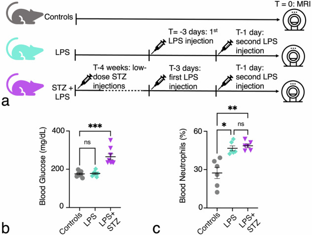
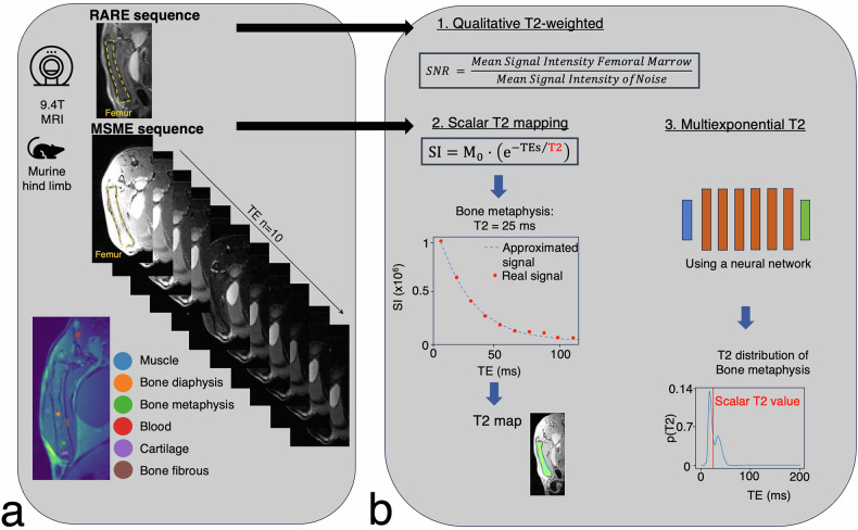
Methods: Using a 9.4-T magnet, T2-weighted and multislice multiecho sequences were applied to evaluate bone marrow in female C57BL/6J mice divided into three groups: (1) controls; (2) lipopolysaccharide-induced acute inflammation (LPS); and (3) streptozotocin (STZ)- and LPS-induced diabetic inflammation (STZ + LPS). T2 relaxation times and their distributions with scalar mapping and model-informed machine learning (MIML) were analyzed. Correlations with histological iron levels and blood neutrophil counts were assessed.
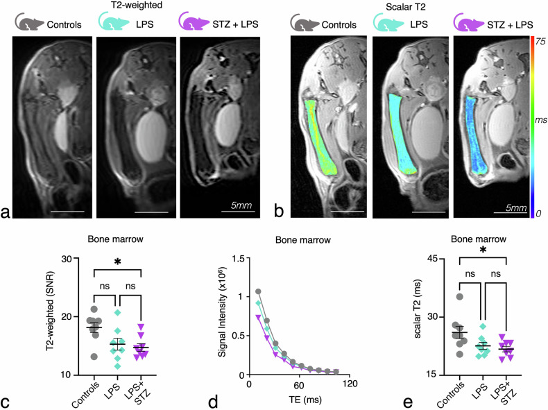
Results: T2-weighted imaging showed a reduced signal-to-noise ratio in inflamed bone marrow (p = 0.034). Scalar T2 mapping identified decreased T2 relaxation times (p = 0.042), moderately correlating with neutrophil counts (ρ = 0.027) and iron levels (ρ = 0.016). MIML-enhanced T2 distribution analysis exhibited superior sensitivity than scalar T2 mapping, revealing significant reductions in the first T2 distribution peak (p = 0.0025), which strongly correlated with neutrophil counts (ρ = 0.0016) and iron sequestration (ρ = 0.0002). Histology confirmed elevated iron deposits in inflamed marrow, aligning with systemic inflammation.
Conclusion: Combining T2-weighted imaging, scalar T2 mapping, and MIML-enhanced T2 distribution analysis offers complementary insights into inflammation-induced bone marrow remodeling. T2 distribution analysis emerged as a more sensitive tool for detecting microstructural changes, such as iron sequestration, supporting its potential as a noninvasive biomarker for diagnosing and monitoring inflammatory diseases.
Relevance statement: This study highlights the potential of advanced MRI T2 analysis and machine learning methods for noninvasive detection of inflammation-induced microstructural changes in bone marrow, offering promising diagnostic tools for inflammatory diseases.
Key points: This study investigated inflammation-induced changes in bone marrow with T2 MRI and MIML. MIML outperformed quantitative scalar T2 analysis, increasingly detecting inflammation and iron sequestration in the hematopoietic bone marrow. T2 MRI with MIML analysis could aid in the early diagnosis and management of inflammatory diseases.

 求助内容:
求助内容: 应助结果提醒方式:


