Theresa Urban, Florian T Gassert, Manuela Frank, Rafael Schick, Henriette Bast, Jannis Bodden, Alexander W Marka, Lisa Steinhelfer, Manuel Steinhardt, Andreas Sauter, Alexander Fingerle, Gregor S Zimmermann, Thomas Koehler, Marcus R Makowski, Daniela Pfeiffer, Franz Pfeiffer
{"title":"健康和肺气肿患者吸气和呼气的暗场胸片信号特征。","authors":"Theresa Urban, Florian T Gassert, Manuela Frank, Rafael Schick, Henriette Bast, Jannis Bodden, Alexander W Marka, Lisa Steinhelfer, Manuel Steinhardt, Andreas Sauter, Alexander Fingerle, Gregor S Zimmermann, Thomas Koehler, Marcus R Makowski, Daniela Pfeiffer, Franz Pfeiffer","doi":"10.1186/s41747-025-00578-x","DOIUrl":null,"url":null,"abstract":"<p><strong>Background: </strong>Dark-field chest radiography is sensitive to the lung alveolar structure. We evaluated the change of dark-field signal between inspiration and expiration.</p><p><strong>Methods: </strong>From 2018 to 2020, patients who underwent chest computed tomography (CT) were prospectively enrolled, excluding those with any lung condition besides emphysema visible on CT. Participants were imaged in both inspiration and expiration with a prototype dark-field chest radiography system. We calculated the total dark-field signal ∑DF and the dark-field coefficient ϵ, assumed to be proportional to the total number of alveoli and the alveolar density, respectively.</p><p><strong>Results: </strong>Eighty-eight subjects, aged 64 years ± 11 (mean ± standard deviation), 55 males, were enrolled. Dark-field signal in the lung projection appeared higher in expiration compared to inspiration. Over all participants, ∑DF was higher in inspiration (1.6 × 10<sup>-2</sup> ± 0.4 × 10<sup>-2</sup> m<sup>2</sup>) compared to expiration (1.5 × 10<sup>-2</sup> ± 0.4 m<sup>2</sup>) (p < 0.001), with its expiration-to-inspiration not ratio being different for any emphysema subgroup. The dark-field coefficient ϵ was lower in inspiration (2.3 ± 0.6 m<sup>-1</sup>) compared to expiration (3.1 ± 1.1 m<sup>-1</sup>) (p < 0.001) over all participants. The dark-field coefficient in inspiration and expiration, as well as their ratio, was lower for at least moderate emphysema when compared to the control group (e.g., ϵ = 2.5 ± 1.0 m<sup>-1</sup> for moderate emphysema in expiration versus ϵ = 3.6 ± 0.7 m<sup>-1</sup> for participants without emphysema (p = 0.003).</p><p><strong>Conclusion: </strong>The dark-field signal depends on the breathing state. Differences between breathing states are influenced by emphysema severity.</p><p><strong>Relevance statement: </strong>The patient's breathing state influences the dark-field chest radiograph, potentially impacting its diagnostic value.</p><p><strong>Key points: </strong>Signal characteristics in dark-field chest radiography change between inspiration and expiration. The total dark-field signal decreases slightly from inspiration to expiration, while the dark-field coefficient increases substantially. The ratio of the total dark-field signal between expiration and inspiration is independent of emphysema severity, whereas the ratio of the dark-field coefficient depends on emphysema severity.</p>","PeriodicalId":36926,"journal":{"name":"European Radiology Experimental","volume":"9 1","pages":"40"},"PeriodicalIF":3.6000,"publicationDate":"2025-03-27","publicationTypes":"Journal Article","fieldsOfStudy":null,"isOpenAccess":false,"openAccessPdf":"https://www.ncbi.nlm.nih.gov/pmc/articles/PMC11950489/pdf/","citationCount":"0","resultStr":"{\"title\":\"Dark-field chest radiography signal characteristics in inspiration and expiration in healthy and emphysematous subjects.\",\"authors\":\"Theresa Urban, Florian T Gassert, Manuela Frank, Rafael Schick, Henriette Bast, Jannis Bodden, Alexander W Marka, Lisa Steinhelfer, Manuel Steinhardt, Andreas Sauter, Alexander Fingerle, Gregor S Zimmermann, Thomas Koehler, Marcus R Makowski, Daniela Pfeiffer, Franz Pfeiffer\",\"doi\":\"10.1186/s41747-025-00578-x\",\"DOIUrl\":null,\"url\":null,\"abstract\":\"<p><strong>Background: </strong>Dark-field chest radiography is sensitive to the lung alveolar structure. We evaluated the change of dark-field signal between inspiration and expiration.</p><p><strong>Methods: </strong>From 2018 to 2020, patients who underwent chest computed tomography (CT) were prospectively enrolled, excluding those with any lung condition besides emphysema visible on CT. Participants were imaged in both inspiration and expiration with a prototype dark-field chest radiography system. We calculated the total dark-field signal ∑DF and the dark-field coefficient ϵ, assumed to be proportional to the total number of alveoli and the alveolar density, respectively.</p><p><strong>Results: </strong>Eighty-eight subjects, aged 64 years ± 11 (mean ± standard deviation), 55 males, were enrolled. Dark-field signal in the lung projection appeared higher in expiration compared to inspiration. Over all participants, ∑DF was higher in inspiration (1.6 × 10<sup>-2</sup> ± 0.4 × 10<sup>-2</sup> m<sup>2</sup>) compared to expiration (1.5 × 10<sup>-2</sup> ± 0.4 m<sup>2</sup>) (p < 0.001), with its expiration-to-inspiration not ratio being different for any emphysema subgroup. The dark-field coefficient ϵ was lower in inspiration (2.3 ± 0.6 m<sup>-1</sup>) compared to expiration (3.1 ± 1.1 m<sup>-1</sup>) (p < 0.001) over all participants. The dark-field coefficient in inspiration and expiration, as well as their ratio, was lower for at least moderate emphysema when compared to the control group (e.g., ϵ = 2.5 ± 1.0 m<sup>-1</sup> for moderate emphysema in expiration versus ϵ = 3.6 ± 0.7 m<sup>-1</sup> for participants without emphysema (p = 0.003).</p><p><strong>Conclusion: </strong>The dark-field signal depends on the breathing state. Differences between breathing states are influenced by emphysema severity.</p><p><strong>Relevance statement: </strong>The patient's breathing state influences the dark-field chest radiograph, potentially impacting its diagnostic value.</p><p><strong>Key points: </strong>Signal characteristics in dark-field chest radiography change between inspiration and expiration. The total dark-field signal decreases slightly from inspiration to expiration, while the dark-field coefficient increases substantially. The ratio of the total dark-field signal between expiration and inspiration is independent of emphysema severity, whereas the ratio of the dark-field coefficient depends on emphysema severity.</p>\",\"PeriodicalId\":36926,\"journal\":{\"name\":\"European Radiology Experimental\",\"volume\":\"9 1\",\"pages\":\"40\"},\"PeriodicalIF\":3.6000,\"publicationDate\":\"2025-03-27\",\"publicationTypes\":\"Journal Article\",\"fieldsOfStudy\":null,\"isOpenAccess\":false,\"openAccessPdf\":\"https://www.ncbi.nlm.nih.gov/pmc/articles/PMC11950489/pdf/\",\"citationCount\":\"0\",\"resultStr\":null,\"platform\":\"Semanticscholar\",\"paperid\":null,\"PeriodicalName\":\"European Radiology Experimental\",\"FirstCategoryId\":\"1085\",\"ListUrlMain\":\"https://doi.org/10.1186/s41747-025-00578-x\",\"RegionNum\":0,\"RegionCategory\":null,\"ArticlePicture\":[],\"TitleCN\":null,\"AbstractTextCN\":null,\"PMCID\":null,\"EPubDate\":\"\",\"PubModel\":\"\",\"JCR\":\"Q1\",\"JCRName\":\"RADIOLOGY, NUCLEAR MEDICINE & MEDICAL IMAGING\",\"Score\":null,\"Total\":0}","platform":"Semanticscholar","paperid":null,"PeriodicalName":"European Radiology Experimental","FirstCategoryId":"1085","ListUrlMain":"https://doi.org/10.1186/s41747-025-00578-x","RegionNum":0,"RegionCategory":null,"ArticlePicture":[],"TitleCN":null,"AbstractTextCN":null,"PMCID":null,"EPubDate":"","PubModel":"","JCR":"Q1","JCRName":"RADIOLOGY, NUCLEAR MEDICINE & MEDICAL IMAGING","Score":null,"Total":0}
Dark-field chest radiography signal characteristics in inspiration and expiration in healthy and emphysematous subjects.
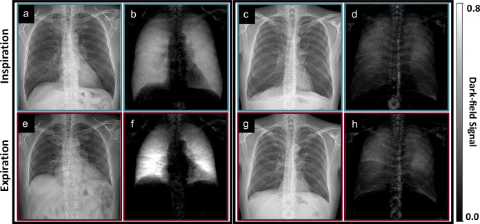
Background: Dark-field chest radiography is sensitive to the lung alveolar structure. We evaluated the change of dark-field signal between inspiration and expiration.
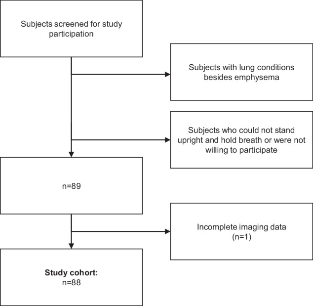
Methods: From 2018 to 2020, patients who underwent chest computed tomography (CT) were prospectively enrolled, excluding those with any lung condition besides emphysema visible on CT. Participants were imaged in both inspiration and expiration with a prototype dark-field chest radiography system. We calculated the total dark-field signal ∑DF and the dark-field coefficient ϵ, assumed to be proportional to the total number of alveoli and the alveolar density, respectively.
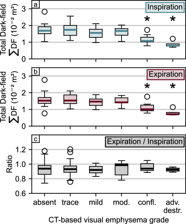
Results: Eighty-eight subjects, aged 64 years ± 11 (mean ± standard deviation), 55 males, were enrolled. Dark-field signal in the lung projection appeared higher in expiration compared to inspiration. Over all participants, ∑DF was higher in inspiration (1.6 × 10⁻² ± 0.4 × 10⁻² m2) compared to expiration (1.5 × 10⁻² ± 0.4 m2) (p < 0.001), with its expiration-to-inspiration not ratio being different for any emphysema subgroup. The dark-field coefficient ϵ was lower in inspiration (2.3 ± 0.6 m-1) compared to expiration (3.1 ± 1.1 m-1) (p < 0.001) over all participants. The dark-field coefficient in inspiration and expiration, as well as their ratio, was lower for at least moderate emphysema when compared to the control group (e.g., ϵ = 2.5 ± 1.0 m-1 for moderate emphysema in expiration versus ϵ = 3.6 ± 0.7 m-1 for participants without emphysema (p = 0.003).
Conclusion: The dark-field signal depends on the breathing state. Differences between breathing states are influenced by emphysema severity.
Relevance statement: The patient's breathing state influences the dark-field chest radiograph, potentially impacting its diagnostic value.
Key points: Signal characteristics in dark-field chest radiography change between inspiration and expiration. The total dark-field signal decreases slightly from inspiration to expiration, while the dark-field coefficient increases substantially. The ratio of the total dark-field signal between expiration and inspiration is independent of emphysema severity, whereas the ratio of the dark-field coefficient depends on emphysema severity.

 求助内容:
求助内容: 应助结果提醒方式:


