Marcio Alex Barros Gomes, Camilla Christian Gomes Moura, Lucas Raineri Capeletti, Gustavo Silva Chaves, Daniel de Almeida Decurcio, Monise de Paula Rodrigues, Carlos José Soares
{"title":"玻璃纤维桩技术去除对牙根牙本质缺失、根管排列和根管穿孔的影响。","authors":"Marcio Alex Barros Gomes, Camilla Christian Gomes Moura, Lucas Raineri Capeletti, Gustavo Silva Chaves, Daniel de Almeida Decurcio, Monise de Paula Rodrigues, Carlos José Soares","doi":"10.1590/0103-644020235851","DOIUrl":null,"url":null,"abstract":"<p><p>Root canal retreatment and fracture of fiberglass posts (FGP) can require the FGP removal. The aim of this study was to evaluate the effect of the FGP removal protocol on the time for FGP removal (min), the root dentin removed (mm3), the angle alteration of the root canal alignment (o), and the root perforation occurrence using cone-beam computed tomography (CBCT) analysis. Thirty extracted maxillary molars were randomly assigned to 3 groups (n = 10): Mic-Ult, FGP removed using ultrasonic inserts under microscopic magnification; Mic-DiB, FGP removed using diamond burs under microscopic magnification, and Endo-G, FGP removed using Endo-G. CBCTs were made after access opening and after FGP removal. The volume of root dentin, the root canal alignment and root perforation were performed using InVesalius and Mimics softwares. Root canal angle alteration after FGP removal was performed using ImageJ software. The time for FGP removal was calculated in min. Data were analysed by one-way analysis of variance was followed by Tukey HSD test (α = 0.05). Endo-G resulted in significantly lower root dentin removal (p< .001), and significantly less time than Mic-Ult and Mic-DiB (p< .001). Mic-DiB had 1 and Mic-Ult had 2 root dentin perforations. FGP post removal using Mic-Ult and Mic-DiB exhibited significantly greater alteration in the root canal alignment than that using Endo-G (p< .001). FGP removal using Endo-G exhibited better preservation of the original canal alignment, saved root dentin structure, and also required less time compared to FGP post removal using Mic-DiB or Mic-Ult under microscopic magnification.</p>","PeriodicalId":101363,"journal":{"name":"Brazilian dental journal","volume":"35 ","pages":"e235851"},"PeriodicalIF":0.0000,"publicationDate":"2024-12-06","publicationTypes":"Journal Article","fieldsOfStudy":null,"isOpenAccess":false,"openAccessPdf":"https://www.ncbi.nlm.nih.gov/pmc/articles/PMC11653786/pdf/","citationCount":"0","resultStr":"{\"title\":\"Effect of Fiberglass Post Technique Removal on the Root Dentin Loss, the Root Canal Alignment and the Root Perforation Occurrence.\",\"authors\":\"Marcio Alex Barros Gomes, Camilla Christian Gomes Moura, Lucas Raineri Capeletti, Gustavo Silva Chaves, Daniel de Almeida Decurcio, Monise de Paula Rodrigues, Carlos José Soares\",\"doi\":\"10.1590/0103-644020235851\",\"DOIUrl\":null,\"url\":null,\"abstract\":\"<p><p>Root canal retreatment and fracture of fiberglass posts (FGP) can require the FGP removal. The aim of this study was to evaluate the effect of the FGP removal protocol on the time for FGP removal (min), the root dentin removed (mm3), the angle alteration of the root canal alignment (o), and the root perforation occurrence using cone-beam computed tomography (CBCT) analysis. Thirty extracted maxillary molars were randomly assigned to 3 groups (n = 10): Mic-Ult, FGP removed using ultrasonic inserts under microscopic magnification; Mic-DiB, FGP removed using diamond burs under microscopic magnification, and Endo-G, FGP removed using Endo-G. CBCTs were made after access opening and after FGP removal. The volume of root dentin, the root canal alignment and root perforation were performed using InVesalius and Mimics softwares. Root canal angle alteration after FGP removal was performed using ImageJ software. The time for FGP removal was calculated in min. Data were analysed by one-way analysis of variance was followed by Tukey HSD test (α = 0.05). Endo-G resulted in significantly lower root dentin removal (p< .001), and significantly less time than Mic-Ult and Mic-DiB (p< .001). Mic-DiB had 1 and Mic-Ult had 2 root dentin perforations. FGP post removal using Mic-Ult and Mic-DiB exhibited significantly greater alteration in the root canal alignment than that using Endo-G (p< .001). FGP removal using Endo-G exhibited better preservation of the original canal alignment, saved root dentin structure, and also required less time compared to FGP post removal using Mic-DiB or Mic-Ult under microscopic magnification.</p>\",\"PeriodicalId\":101363,\"journal\":{\"name\":\"Brazilian dental journal\",\"volume\":\"35 \",\"pages\":\"e235851\"},\"PeriodicalIF\":0.0000,\"publicationDate\":\"2024-12-06\",\"publicationTypes\":\"Journal Article\",\"fieldsOfStudy\":null,\"isOpenAccess\":false,\"openAccessPdf\":\"https://www.ncbi.nlm.nih.gov/pmc/articles/PMC11653786/pdf/\",\"citationCount\":\"0\",\"resultStr\":null,\"platform\":\"Semanticscholar\",\"paperid\":null,\"PeriodicalName\":\"Brazilian dental journal\",\"FirstCategoryId\":\"1085\",\"ListUrlMain\":\"https://doi.org/10.1590/0103-644020235851\",\"RegionNum\":0,\"RegionCategory\":null,\"ArticlePicture\":[],\"TitleCN\":null,\"AbstractTextCN\":null,\"PMCID\":null,\"EPubDate\":\"2024/1/1 0:00:00\",\"PubModel\":\"eCollection\",\"JCR\":\"\",\"JCRName\":\"\",\"Score\":null,\"Total\":0}","platform":"Semanticscholar","paperid":null,"PeriodicalName":"Brazilian dental journal","FirstCategoryId":"1085","ListUrlMain":"https://doi.org/10.1590/0103-644020235851","RegionNum":0,"RegionCategory":null,"ArticlePicture":[],"TitleCN":null,"AbstractTextCN":null,"PMCID":null,"EPubDate":"2024/1/1 0:00:00","PubModel":"eCollection","JCR":"","JCRName":"","Score":null,"Total":0}
Effect of Fiberglass Post Technique Removal on the Root Dentin Loss, the Root Canal Alignment and the Root Perforation Occurrence.
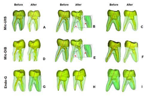
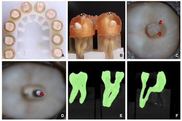
Root canal retreatment and fracture of fiberglass posts (FGP) can require the FGP removal. The aim of this study was to evaluate the effect of the FGP removal protocol on the time for FGP removal (min), the root dentin removed (mm3), the angle alteration of the root canal alignment (o), and the root perforation occurrence using cone-beam computed tomography (CBCT) analysis. Thirty extracted maxillary molars were randomly assigned to 3 groups (n = 10): Mic-Ult, FGP removed using ultrasonic inserts under microscopic magnification; Mic-DiB, FGP removed using diamond burs under microscopic magnification, and Endo-G, FGP removed using Endo-G. CBCTs were made after access opening and after FGP removal. The volume of root dentin, the root canal alignment and root perforation were performed using InVesalius and Mimics softwares. Root canal angle alteration after FGP removal was performed using ImageJ software. The time for FGP removal was calculated in min. Data were analysed by one-way analysis of variance was followed by Tukey HSD test (α = 0.05). Endo-G resulted in significantly lower root dentin removal (p< .001), and significantly less time than Mic-Ult and Mic-DiB (p< .001). Mic-DiB had 1 and Mic-Ult had 2 root dentin perforations. FGP post removal using Mic-Ult and Mic-DiB exhibited significantly greater alteration in the root canal alignment than that using Endo-G (p< .001). FGP removal using Endo-G exhibited better preservation of the original canal alignment, saved root dentin structure, and also required less time compared to FGP post removal using Mic-DiB or Mic-Ult under microscopic magnification.

 求助内容:
求助内容: 应助结果提醒方式:


