Jaromír Vašíček, Andrej Baláži, Mária Tirpáková, Marián Tomka, Peter Chrenek
{"title":"兔干/祖细胞低温保存后细胞内宏微量元素组成的变化。","authors":"Jaromír Vašíček, Andrej Baláži, Mária Tirpáková, Marián Tomka, Peter Chrenek","doi":"10.3390/jdb13010006","DOIUrl":null,"url":null,"abstract":"<p><p>Cryopreservation is a widely used method for the long-term preservation of reproductive or somatic cells. It is known that this storage method may negatively affect cell viability, proliferation, differentiation, etc. However, there is a lack of information about whether cryostorage can alter the content of intracellular minerals. Therefore, we focused this study on the analysis of the mineral composition of living cells before and after long-term cold storage. Briefly, three different primary cell lines were established from rabbits as follows: endothelial progenitor cells from peripheral blood (EPCs), endothelial progenitor cells from bone marrow (BEPCs), and mesenchymal stem cells from adipose tissue (AT-MSCs), which were cultured until passage 3 prior to cryopreservation in liquid nitrogen. Samples from freshly cultured and frozen-thawed cells were mineralized and analyzed using inductively coupled plasma-optical emission spectroscopy (ICP-OES) for the content of minerals (macro: Ca, Na, K, and Mg, and micro: Zn, Fe, Cu, Al, Co, Mn, Sr, and Ni). After cryopreservation, we found significantly decreased content of K in frozen-thawed EPCs (<i>p</i> < 0.01) and BEPCs (<i>p</i> < 0.0001) and Ca in AT-MSCs (<i>p</i> < 0.05), while Na was increased in frozen-thawed BEPCs (<i>p</i> < 0.05). Concentrations of Fe and Al were reduced significantly in frozen-thawed EPCs (both <i>p</i> < 0.0001) and AT-MSCs (<i>p</i> < 0.001 and <i>p</i> < 0.0001, respectively). On the contrary, Fe and Al were elevated in frozen-thawed BEPCs (<i>p</i> < 0.0001 and <i>p</i> < 0.01, respectively) together with Ni (<i>p</i> < 0.0001). In addition, decreased Zn (<i>p</i> < 0.05) was observed in cryopreserved AT-MSCs. In conclusion, the ICP-OES technique might be used to analyze the basic elemental composition of animal cells in fresh or frozen-thawed conditions. Nevertheless, additional studies are needed to reveal the possible impact of cryopreservation on cell fate by changing the content of intracellular minerals.</p>","PeriodicalId":15563,"journal":{"name":"Journal of Developmental Biology","volume":"13 1","pages":""},"PeriodicalIF":2.5000,"publicationDate":"2025-02-21","publicationTypes":"Journal Article","fieldsOfStudy":null,"isOpenAccess":false,"openAccessPdf":"https://www.ncbi.nlm.nih.gov/pmc/articles/PMC11942849/pdf/","citationCount":"0","resultStr":"{\"title\":\"Changes in the Intracellular Composition of Macro and Microminerals After Cryopreservation of the Rabbit Stem/Progenitor Cells.\",\"authors\":\"Jaromír Vašíček, Andrej Baláži, Mária Tirpáková, Marián Tomka, Peter Chrenek\",\"doi\":\"10.3390/jdb13010006\",\"DOIUrl\":null,\"url\":null,\"abstract\":\"<p><p>Cryopreservation is a widely used method for the long-term preservation of reproductive or somatic cells. It is known that this storage method may negatively affect cell viability, proliferation, differentiation, etc. However, there is a lack of information about whether cryostorage can alter the content of intracellular minerals. Therefore, we focused this study on the analysis of the mineral composition of living cells before and after long-term cold storage. Briefly, three different primary cell lines were established from rabbits as follows: endothelial progenitor cells from peripheral blood (EPCs), endothelial progenitor cells from bone marrow (BEPCs), and mesenchymal stem cells from adipose tissue (AT-MSCs), which were cultured until passage 3 prior to cryopreservation in liquid nitrogen. Samples from freshly cultured and frozen-thawed cells were mineralized and analyzed using inductively coupled plasma-optical emission spectroscopy (ICP-OES) for the content of minerals (macro: Ca, Na, K, and Mg, and micro: Zn, Fe, Cu, Al, Co, Mn, Sr, and Ni). After cryopreservation, we found significantly decreased content of K in frozen-thawed EPCs (<i>p</i> < 0.01) and BEPCs (<i>p</i> < 0.0001) and Ca in AT-MSCs (<i>p</i> < 0.05), while Na was increased in frozen-thawed BEPCs (<i>p</i> < 0.05). Concentrations of Fe and Al were reduced significantly in frozen-thawed EPCs (both <i>p</i> < 0.0001) and AT-MSCs (<i>p</i> < 0.001 and <i>p</i> < 0.0001, respectively). On the contrary, Fe and Al were elevated in frozen-thawed BEPCs (<i>p</i> < 0.0001 and <i>p</i> < 0.01, respectively) together with Ni (<i>p</i> < 0.0001). In addition, decreased Zn (<i>p</i> < 0.05) was observed in cryopreserved AT-MSCs. In conclusion, the ICP-OES technique might be used to analyze the basic elemental composition of animal cells in fresh or frozen-thawed conditions. Nevertheless, additional studies are needed to reveal the possible impact of cryopreservation on cell fate by changing the content of intracellular minerals.</p>\",\"PeriodicalId\":15563,\"journal\":{\"name\":\"Journal of Developmental Biology\",\"volume\":\"13 1\",\"pages\":\"\"},\"PeriodicalIF\":2.5000,\"publicationDate\":\"2025-02-21\",\"publicationTypes\":\"Journal Article\",\"fieldsOfStudy\":null,\"isOpenAccess\":false,\"openAccessPdf\":\"https://www.ncbi.nlm.nih.gov/pmc/articles/PMC11942849/pdf/\",\"citationCount\":\"0\",\"resultStr\":null,\"platform\":\"Semanticscholar\",\"paperid\":null,\"PeriodicalName\":\"Journal of Developmental Biology\",\"FirstCategoryId\":\"1085\",\"ListUrlMain\":\"https://doi.org/10.3390/jdb13010006\",\"RegionNum\":0,\"RegionCategory\":null,\"ArticlePicture\":[],\"TitleCN\":null,\"AbstractTextCN\":null,\"PMCID\":null,\"EPubDate\":\"\",\"PubModel\":\"\",\"JCR\":\"Q3\",\"JCRName\":\"DEVELOPMENTAL BIOLOGY\",\"Score\":null,\"Total\":0}","platform":"Semanticscholar","paperid":null,"PeriodicalName":"Journal of Developmental Biology","FirstCategoryId":"1085","ListUrlMain":"https://doi.org/10.3390/jdb13010006","RegionNum":0,"RegionCategory":null,"ArticlePicture":[],"TitleCN":null,"AbstractTextCN":null,"PMCID":null,"EPubDate":"","PubModel":"","JCR":"Q3","JCRName":"DEVELOPMENTAL BIOLOGY","Score":null,"Total":0}
Changes in the Intracellular Composition of Macro and Microminerals After Cryopreservation of the Rabbit Stem/Progenitor Cells.
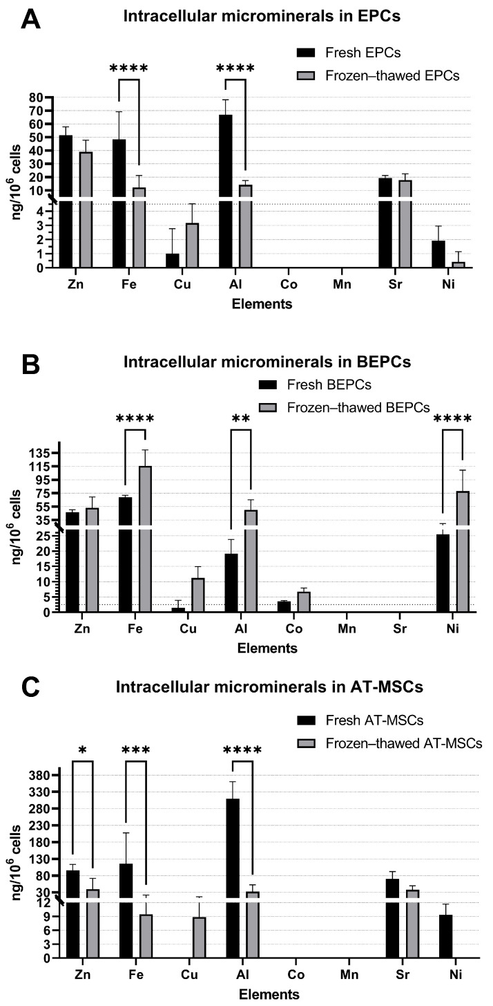
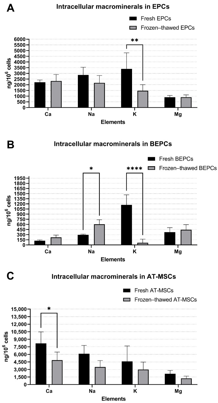
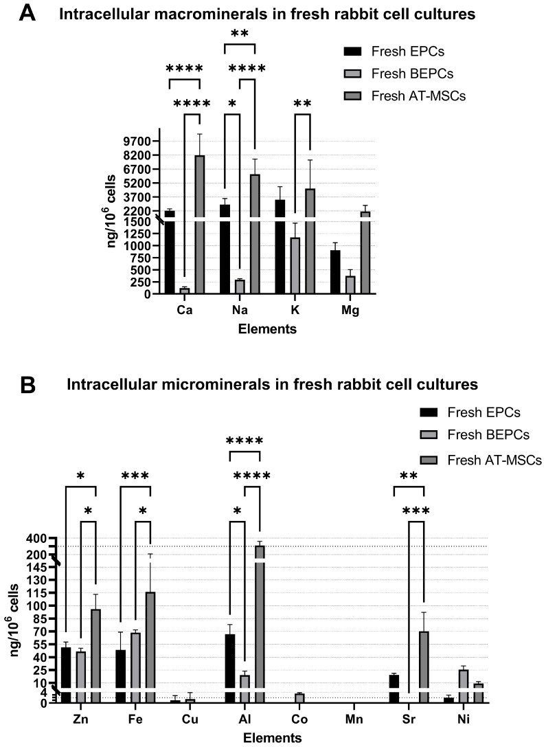
Cryopreservation is a widely used method for the long-term preservation of reproductive or somatic cells. It is known that this storage method may negatively affect cell viability, proliferation, differentiation, etc. However, there is a lack of information about whether cryostorage can alter the content of intracellular minerals. Therefore, we focused this study on the analysis of the mineral composition of living cells before and after long-term cold storage. Briefly, three different primary cell lines were established from rabbits as follows: endothelial progenitor cells from peripheral blood (EPCs), endothelial progenitor cells from bone marrow (BEPCs), and mesenchymal stem cells from adipose tissue (AT-MSCs), which were cultured until passage 3 prior to cryopreservation in liquid nitrogen. Samples from freshly cultured and frozen-thawed cells were mineralized and analyzed using inductively coupled plasma-optical emission spectroscopy (ICP-OES) for the content of minerals (macro: Ca, Na, K, and Mg, and micro: Zn, Fe, Cu, Al, Co, Mn, Sr, and Ni). After cryopreservation, we found significantly decreased content of K in frozen-thawed EPCs (p < 0.01) and BEPCs (p < 0.0001) and Ca in AT-MSCs (p < 0.05), while Na was increased in frozen-thawed BEPCs (p < 0.05). Concentrations of Fe and Al were reduced significantly in frozen-thawed EPCs (both p < 0.0001) and AT-MSCs (p < 0.001 and p < 0.0001, respectively). On the contrary, Fe and Al were elevated in frozen-thawed BEPCs (p < 0.0001 and p < 0.01, respectively) together with Ni (p < 0.0001). In addition, decreased Zn (p < 0.05) was observed in cryopreserved AT-MSCs. In conclusion, the ICP-OES technique might be used to analyze the basic elemental composition of animal cells in fresh or frozen-thawed conditions. Nevertheless, additional studies are needed to reveal the possible impact of cryopreservation on cell fate by changing the content of intracellular minerals.
期刊介绍:
The Journal of Developmental Biology (ISSN 2221-3759) is an international, peer-reviewed, quick-refereeing, open access journal, which publishes reviews, research papers and communications on the development of multicellular organisms at the molecule, cell, tissue, organ and whole organism levels. Our aim is to encourage researchers to effortlessly publish their new findings or concepts rapidly in an open access medium, overseen by their peers. There is no restriction on the length of the papers; the full experimental details must be provided so that the results can be reproduced. Electronic files regarding the full details of the experimental procedure, if unable to be published in a normal way, can be deposited as supplementary material. Journal of Developmental Biology focuses on: -Development mechanisms and genetics -Cell differentiation -Embryonal development -Tissue/organism growth -Metamorphosis and regeneration of the organisms. It involves many biological fields, such as Molecular biology, Genetics, Physiology, Cell biology, Anatomy, Embryology, Cancer research, Neurobiology, Immunology, Ecology, Evolutionary biology.

 求助内容:
求助内容: 应助结果提醒方式:


