Sruthi Kallayil Suresh, Girija Venkatesh Arishinagodi, Mallanagouda B Patil
{"title":"低水平激光治疗对富血小板纤维蛋白影响的显微评价:体外研究。","authors":"Sruthi Kallayil Suresh, Girija Venkatesh Arishinagodi, Mallanagouda B Patil","doi":"10.4103/jisp.jisp_534_23","DOIUrl":null,"url":null,"abstract":"<p><strong>Background: </strong>Platelet-rich fibrin (PRF) serves as a natural fibrin-based biomaterial scaffold, facilitating cellular attachment. Low-level laser therapy (LLLT) may influence PRF properties through its stimulatory effects.</p><p><strong>Aim: </strong>The study aimed to evaluate the effect of LLLT on PRF architectural changes and platelet concentrate values.</p><p><strong>Materials and methods: </strong>Thirty-four samples from seventeen subjects were included in the study, with 20 mL of blood collected from each participant. Blood was distributed into two 10 mL vacutainer tubes: one served as a control for baseline platelet and leukocyte counts, whereas the other was used for PRF preparation. The PRF samples were vertically cut into two equal parts and divided into two groups: Group I (without laser treatment) and Group II (treated with LLLT). Histological preparations were performed for all samples. The mean and standard deviation of platelet and leukocyte counts in Groups I and II were analyzed using the Statistical Package for the Social Sciences (SPSS) version 17.1. Analysis of variance (ANOVA) and <i>post hoc</i> tests were employed for comparisons.</p><p><strong>Results: </strong>Platelet and leukocyte values showed statistically significant differences, with greater cellular entrapment in Group II compared to Group I. The residual serum cell count in both groups was lower than the whole blood cell count. Microscopically, Group I displayed dense, thick fibrils with increased spacing distally, whereas Group II exhibited thinner fibrils with gradual density changes toward the distal end.</p><p><strong>Conclusion: </strong>PRF obtained in both groups was similar in size. Laser irradiation altered the fibrin architecture and enhanced platelet and leukocyte entrapment in PRF.</p>","PeriodicalId":15890,"journal":{"name":"Journal of Indian Society of Periodontology","volume":"28 5","pages":"529-532"},"PeriodicalIF":0.0000,"publicationDate":"2024-09-01","publicationTypes":"Journal Article","fieldsOfStudy":null,"isOpenAccess":false,"openAccessPdf":"https://www.ncbi.nlm.nih.gov/pmc/articles/PMC11932570/pdf/","citationCount":"0","resultStr":"{\"title\":\"Microscopic evaluation of the effect of low-level laser therapy on platelet-rich fibrin: An <i>in vitro</i> study.\",\"authors\":\"Sruthi Kallayil Suresh, Girija Venkatesh Arishinagodi, Mallanagouda B Patil\",\"doi\":\"10.4103/jisp.jisp_534_23\",\"DOIUrl\":null,\"url\":null,\"abstract\":\"<p><strong>Background: </strong>Platelet-rich fibrin (PRF) serves as a natural fibrin-based biomaterial scaffold, facilitating cellular attachment. Low-level laser therapy (LLLT) may influence PRF properties through its stimulatory effects.</p><p><strong>Aim: </strong>The study aimed to evaluate the effect of LLLT on PRF architectural changes and platelet concentrate values.</p><p><strong>Materials and methods: </strong>Thirty-four samples from seventeen subjects were included in the study, with 20 mL of blood collected from each participant. Blood was distributed into two 10 mL vacutainer tubes: one served as a control for baseline platelet and leukocyte counts, whereas the other was used for PRF preparation. The PRF samples were vertically cut into two equal parts and divided into two groups: Group I (without laser treatment) and Group II (treated with LLLT). Histological preparations were performed for all samples. The mean and standard deviation of platelet and leukocyte counts in Groups I and II were analyzed using the Statistical Package for the Social Sciences (SPSS) version 17.1. Analysis of variance (ANOVA) and <i>post hoc</i> tests were employed for comparisons.</p><p><strong>Results: </strong>Platelet and leukocyte values showed statistically significant differences, with greater cellular entrapment in Group II compared to Group I. The residual serum cell count in both groups was lower than the whole blood cell count. Microscopically, Group I displayed dense, thick fibrils with increased spacing distally, whereas Group II exhibited thinner fibrils with gradual density changes toward the distal end.</p><p><strong>Conclusion: </strong>PRF obtained in both groups was similar in size. Laser irradiation altered the fibrin architecture and enhanced platelet and leukocyte entrapment in PRF.</p>\",\"PeriodicalId\":15890,\"journal\":{\"name\":\"Journal of Indian Society of Periodontology\",\"volume\":\"28 5\",\"pages\":\"529-532\"},\"PeriodicalIF\":0.0000,\"publicationDate\":\"2024-09-01\",\"publicationTypes\":\"Journal Article\",\"fieldsOfStudy\":null,\"isOpenAccess\":false,\"openAccessPdf\":\"https://www.ncbi.nlm.nih.gov/pmc/articles/PMC11932570/pdf/\",\"citationCount\":\"0\",\"resultStr\":null,\"platform\":\"Semanticscholar\",\"paperid\":null,\"PeriodicalName\":\"Journal of Indian Society of Periodontology\",\"FirstCategoryId\":\"1085\",\"ListUrlMain\":\"https://doi.org/10.4103/jisp.jisp_534_23\",\"RegionNum\":0,\"RegionCategory\":null,\"ArticlePicture\":[],\"TitleCN\":null,\"AbstractTextCN\":null,\"PMCID\":null,\"EPubDate\":\"2025/2/26 0:00:00\",\"PubModel\":\"Epub\",\"JCR\":\"Q2\",\"JCRName\":\"Dentistry\",\"Score\":null,\"Total\":0}","platform":"Semanticscholar","paperid":null,"PeriodicalName":"Journal of Indian Society of Periodontology","FirstCategoryId":"1085","ListUrlMain":"https://doi.org/10.4103/jisp.jisp_534_23","RegionNum":0,"RegionCategory":null,"ArticlePicture":[],"TitleCN":null,"AbstractTextCN":null,"PMCID":null,"EPubDate":"2025/2/26 0:00:00","PubModel":"Epub","JCR":"Q2","JCRName":"Dentistry","Score":null,"Total":0}
Microscopic evaluation of the effect of low-level laser therapy on platelet-rich fibrin: An in vitro study.
Background: Platelet-rich fibrin (PRF) serves as a natural fibrin-based biomaterial scaffold, facilitating cellular attachment. Low-level laser therapy (LLLT) may influence PRF properties through its stimulatory effects.
Aim: The study aimed to evaluate the effect of LLLT on PRF architectural changes and platelet concentrate values.
Materials and methods: Thirty-four samples from seventeen subjects were included in the study, with 20 mL of blood collected from each participant. Blood was distributed into two 10 mL vacutainer tubes: one served as a control for baseline platelet and leukocyte counts, whereas the other was used for PRF preparation. The PRF samples were vertically cut into two equal parts and divided into two groups: Group I (without laser treatment) and Group II (treated with LLLT). Histological preparations were performed for all samples. The mean and standard deviation of platelet and leukocyte counts in Groups I and II were analyzed using the Statistical Package for the Social Sciences (SPSS) version 17.1. Analysis of variance (ANOVA) and post hoc tests were employed for comparisons.
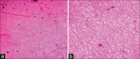
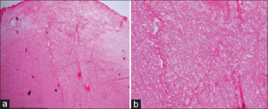
Results: Platelet and leukocyte values showed statistically significant differences, with greater cellular entrapment in Group II compared to Group I. The residual serum cell count in both groups was lower than the whole blood cell count. Microscopically, Group I displayed dense, thick fibrils with increased spacing distally, whereas Group II exhibited thinner fibrils with gradual density changes toward the distal end.
Conclusion: PRF obtained in both groups was similar in size. Laser irradiation altered the fibrin architecture and enhanced platelet and leukocyte entrapment in PRF.
期刊介绍:
The Journal of Indian Society of Periodontology publishes original scientific articles to support practice , education and research in the dental specialty of periodontology and oral implantology. Journal of Indian Society of Periodontology (JISP), is the official publication of the Society and is managed and brought out by the Editor of the society. The journal is published Bimonthly with special issues being brought out for specific occasions. The ISP had a bulletin as its publication for a large number of years and was enhanced as a Journal a few years ago

 求助内容:
求助内容: 应助结果提醒方式:


