Caroline G G Beltran, Jurgen Kriel, Stefan M Botha, Margaret B Nolan, Alessandro Ciccarelli, Ben Loos, Maximiliano G Gutierrez, Gerhard Walzl
{"title":"相关三维成像方法分析易感小鼠结核分枝杆菌病变结构。","authors":"Caroline G G Beltran, Jurgen Kriel, Stefan M Botha, Margaret B Nolan, Alessandro Ciccarelli, Ben Loos, Maximiliano G Gutierrez, Gerhard Walzl","doi":"10.1242/dmm.052185","DOIUrl":null,"url":null,"abstract":"<p><p>Tuberculosis (TB) is characterized by the formation of heterogeneous, immune-rich granulomas in the lungs. Host and pathogen factors contribute to this heterogeneity, but the molecular and cellular drivers of granuloma diversity remain inadequately understood owing to limitations in experimental techniques. In this study, we developed an approach that combines passive CLARITY (PACT)-based clearing with light-sheet fluorescence microscopy to visualize lesion architecture and lung involvement in Mycobacterium tuberculosis-infected C3HeB/FeJ mice. Three-dimensional rendering of post-mortem lungs revealed critical architectural features in lesion development that traditional thin-section imaging could not detect. Wild-type M. tuberculosis infection resulted in organized granulomas, with median sizes increasing to 3.74×108 µm3 and occupying ∼10% of the total lung volume by day 70 post-infection. In contrast, infection with the avirulent ESX-1 deletion mutant strain resulted in diffuse and sparsely organized CD11b recruitment (median size of 8.22×107 µm3), primarily located in the lung periphery and minimally involving the airways (0.23% of the total lung space). Additionally, we present a method for volumetric correlative light and electron microscopy, enabling tracking of individual immune cell populations within granulomas.</p>","PeriodicalId":11144,"journal":{"name":"Disease Models & Mechanisms","volume":"18 9","pages":""},"PeriodicalIF":3.3000,"publicationDate":"2025-09-01","publicationTypes":"Journal Article","fieldsOfStudy":null,"isOpenAccess":false,"openAccessPdf":"https://www.ncbi.nlm.nih.gov/pmc/articles/PMC11972079/pdf/","citationCount":"0","resultStr":"{\"title\":\"Correlative 3D imaging method for analysing lesion architecture in susceptible mice infected with Mycobacterium tuberculosis.\",\"authors\":\"Caroline G G Beltran, Jurgen Kriel, Stefan M Botha, Margaret B Nolan, Alessandro Ciccarelli, Ben Loos, Maximiliano G Gutierrez, Gerhard Walzl\",\"doi\":\"10.1242/dmm.052185\",\"DOIUrl\":null,\"url\":null,\"abstract\":\"<p><p>Tuberculosis (TB) is characterized by the formation of heterogeneous, immune-rich granulomas in the lungs. Host and pathogen factors contribute to this heterogeneity, but the molecular and cellular drivers of granuloma diversity remain inadequately understood owing to limitations in experimental techniques. In this study, we developed an approach that combines passive CLARITY (PACT)-based clearing with light-sheet fluorescence microscopy to visualize lesion architecture and lung involvement in Mycobacterium tuberculosis-infected C3HeB/FeJ mice. Three-dimensional rendering of post-mortem lungs revealed critical architectural features in lesion development that traditional thin-section imaging could not detect. Wild-type M. tuberculosis infection resulted in organized granulomas, with median sizes increasing to 3.74×108 µm3 and occupying ∼10% of the total lung volume by day 70 post-infection. In contrast, infection with the avirulent ESX-1 deletion mutant strain resulted in diffuse and sparsely organized CD11b recruitment (median size of 8.22×107 µm3), primarily located in the lung periphery and minimally involving the airways (0.23% of the total lung space). Additionally, we present a method for volumetric correlative light and electron microscopy, enabling tracking of individual immune cell populations within granulomas.</p>\",\"PeriodicalId\":11144,\"journal\":{\"name\":\"Disease Models & Mechanisms\",\"volume\":\"18 9\",\"pages\":\"\"},\"PeriodicalIF\":3.3000,\"publicationDate\":\"2025-09-01\",\"publicationTypes\":\"Journal Article\",\"fieldsOfStudy\":null,\"isOpenAccess\":false,\"openAccessPdf\":\"https://www.ncbi.nlm.nih.gov/pmc/articles/PMC11972079/pdf/\",\"citationCount\":\"0\",\"resultStr\":null,\"platform\":\"Semanticscholar\",\"paperid\":null,\"PeriodicalName\":\"Disease Models & Mechanisms\",\"FirstCategoryId\":\"3\",\"ListUrlMain\":\"https://doi.org/10.1242/dmm.052185\",\"RegionNum\":3,\"RegionCategory\":\"医学\",\"ArticlePicture\":[],\"TitleCN\":null,\"AbstractTextCN\":null,\"PMCID\":null,\"EPubDate\":\"2025/3/26 0:00:00\",\"PubModel\":\"Epub\",\"JCR\":\"Q2\",\"JCRName\":\"CELL BIOLOGY\",\"Score\":null,\"Total\":0}","platform":"Semanticscholar","paperid":null,"PeriodicalName":"Disease Models & Mechanisms","FirstCategoryId":"3","ListUrlMain":"https://doi.org/10.1242/dmm.052185","RegionNum":3,"RegionCategory":"医学","ArticlePicture":[],"TitleCN":null,"AbstractTextCN":null,"PMCID":null,"EPubDate":"2025/3/26 0:00:00","PubModel":"Epub","JCR":"Q2","JCRName":"CELL BIOLOGY","Score":null,"Total":0}
Correlative 3D imaging method for analysing lesion architecture in susceptible mice infected with Mycobacterium tuberculosis.
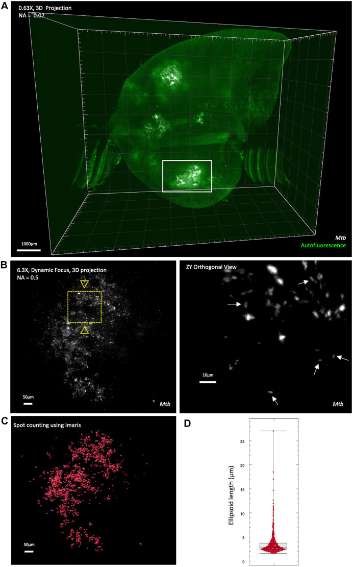
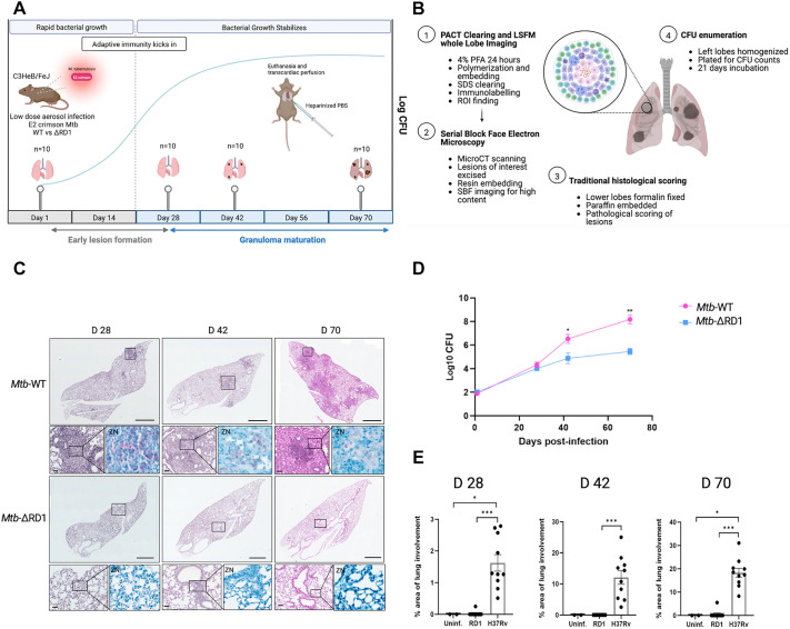
Tuberculosis (TB) is characterized by the formation of heterogeneous, immune-rich granulomas in the lungs. Host and pathogen factors contribute to this heterogeneity, but the molecular and cellular drivers of granuloma diversity remain inadequately understood owing to limitations in experimental techniques. In this study, we developed an approach that combines passive CLARITY (PACT)-based clearing with light-sheet fluorescence microscopy to visualize lesion architecture and lung involvement in Mycobacterium tuberculosis-infected C3HeB/FeJ mice. Three-dimensional rendering of post-mortem lungs revealed critical architectural features in lesion development that traditional thin-section imaging could not detect. Wild-type M. tuberculosis infection resulted in organized granulomas, with median sizes increasing to 3.74×108 µm3 and occupying ∼10% of the total lung volume by day 70 post-infection. In contrast, infection with the avirulent ESX-1 deletion mutant strain resulted in diffuse and sparsely organized CD11b recruitment (median size of 8.22×107 µm3), primarily located in the lung periphery and minimally involving the airways (0.23% of the total lung space). Additionally, we present a method for volumetric correlative light and electron microscopy, enabling tracking of individual immune cell populations within granulomas.
期刊介绍:
Disease Models & Mechanisms (DMM) is an online Open Access journal focusing on the use of model systems to better understand, diagnose and treat human disease.
 求助内容:
求助内容: 应助结果提醒方式:


