Marc G. Sarossy, Sandy Rezk, Dongzhe Li, Xavier Hadoux, Maxime Jannaud, Michael A. Coote, Ghee Soon Ang, Peter van Wijngaarden, Keith R. Martin, Zhichao Wu
{"title":"高分辨率显微镜检测青光眼损害:性能的前瞻性评价。","authors":"Marc G. Sarossy, Sandy Rezk, Dongzhe Li, Xavier Hadoux, Maxime Jannaud, Michael A. Coote, Ghee Soon Ang, Peter van Wijngaarden, Keith R. Martin, Zhichao Wu","doi":"10.1111/ceo.14528","DOIUrl":null,"url":null,"abstract":"<div>\n \n \n <section>\n \n <h3> Background</h3>\n \n <p>To compare the performance of high-resolution microperimetry testing against standard automated perimetry (SAP) for detecting glaucomatous damage seen on optical coherence tomography (OCT) scans.</p>\n </section>\n \n <section>\n \n <h3> Methods</h3>\n \n <p>250 eyes from 200 individuals underwent high-resolution microperimetry testing of a hemifield using a stimulus pattern optimised to sample typical arcuate patterns of visual field loss in glaucoma. SAP was performed using a 24–2 stimulus pattern. The presence of glaucomatous damage in each hemifield was subsequently independently assessed by two graders based on a circumpapillary OCT circle scan, 6×6 mm optic disc-centred OCT volume scan, and 15×15 mm widefield OCT volume scan.</p>\n </section>\n \n <section>\n \n <h3> Results</h3>\n \n <p>The hemifield-based mean total deviation (MTD) and pointwise sensitivity standard deviation (PSD) for high-resolution microperimetry and SAP were both independently associated with the presence of glaucomatous damage in multivariable logistic regression analyses (<i>p</i> ≤ 0.046). Prediction models developed using these two parameters showed significantly higher performance with high-resolution microperimetry compared to SAP (partial area under the receiver operating characteristic curve [pAUC] = 0.86 and 0.75 respectively; <i>p</i> = 0.007). The performance of microperimetry decreased with decreasing resolution when the MTD and PSD values were derived from every second, third, and sixth test locations (pAUC = 0.85 [<i>p</i> = 0.16], 0.85 [<i>p</i> = 0.044] and 0.84 [<i>p</i> < 0.001] respectively), but they were all significantly better than SAP (<i>p</i> ≤ 0.028 for all).</p>\n </section>\n \n <section>\n \n <h3> Conclusions</h3>\n \n <p>High-resolution microperimetry enabled improved performance for detecting glaucomatous damage compared to SAP, highlighting the potential value of fundus-tracking and higher-resolution sampling for the detection of glaucomatous visual field loss.</p>\n </section>\n </div>","PeriodicalId":55253,"journal":{"name":"Clinical and Experimental Ophthalmology","volume":"53 6","pages":"602-610"},"PeriodicalIF":5.6000,"publicationDate":"2025-03-24","publicationTypes":"Journal Article","fieldsOfStudy":null,"isOpenAccess":false,"openAccessPdf":"https://onlinelibrary.wiley.com/doi/epdf/10.1111/ceo.14528","citationCount":"0","resultStr":"{\"title\":\"High-Resolution Microperimetry for Detecting Glaucomatous Damage: A Prospective Evaluation of Performance\",\"authors\":\"Marc G. Sarossy, Sandy Rezk, Dongzhe Li, Xavier Hadoux, Maxime Jannaud, Michael A. Coote, Ghee Soon Ang, Peter van Wijngaarden, Keith R. Martin, Zhichao Wu\",\"doi\":\"10.1111/ceo.14528\",\"DOIUrl\":null,\"url\":null,\"abstract\":\"<div>\\n \\n \\n <section>\\n \\n <h3> Background</h3>\\n \\n <p>To compare the performance of high-resolution microperimetry testing against standard automated perimetry (SAP) for detecting glaucomatous damage seen on optical coherence tomography (OCT) scans.</p>\\n </section>\\n \\n <section>\\n \\n <h3> Methods</h3>\\n \\n <p>250 eyes from 200 individuals underwent high-resolution microperimetry testing of a hemifield using a stimulus pattern optimised to sample typical arcuate patterns of visual field loss in glaucoma. SAP was performed using a 24–2 stimulus pattern. The presence of glaucomatous damage in each hemifield was subsequently independently assessed by two graders based on a circumpapillary OCT circle scan, 6×6 mm optic disc-centred OCT volume scan, and 15×15 mm widefield OCT volume scan.</p>\\n </section>\\n \\n <section>\\n \\n <h3> Results</h3>\\n \\n <p>The hemifield-based mean total deviation (MTD) and pointwise sensitivity standard deviation (PSD) for high-resolution microperimetry and SAP were both independently associated with the presence of glaucomatous damage in multivariable logistic regression analyses (<i>p</i> ≤ 0.046). Prediction models developed using these two parameters showed significantly higher performance with high-resolution microperimetry compared to SAP (partial area under the receiver operating characteristic curve [pAUC] = 0.86 and 0.75 respectively; <i>p</i> = 0.007). The performance of microperimetry decreased with decreasing resolution when the MTD and PSD values were derived from every second, third, and sixth test locations (pAUC = 0.85 [<i>p</i> = 0.16], 0.85 [<i>p</i> = 0.044] and 0.84 [<i>p</i> < 0.001] respectively), but they were all significantly better than SAP (<i>p</i> ≤ 0.028 for all).</p>\\n </section>\\n \\n <section>\\n \\n <h3> Conclusions</h3>\\n \\n <p>High-resolution microperimetry enabled improved performance for detecting glaucomatous damage compared to SAP, highlighting the potential value of fundus-tracking and higher-resolution sampling for the detection of glaucomatous visual field loss.</p>\\n </section>\\n </div>\",\"PeriodicalId\":55253,\"journal\":{\"name\":\"Clinical and Experimental Ophthalmology\",\"volume\":\"53 6\",\"pages\":\"602-610\"},\"PeriodicalIF\":5.6000,\"publicationDate\":\"2025-03-24\",\"publicationTypes\":\"Journal Article\",\"fieldsOfStudy\":null,\"isOpenAccess\":false,\"openAccessPdf\":\"https://onlinelibrary.wiley.com/doi/epdf/10.1111/ceo.14528\",\"citationCount\":\"0\",\"resultStr\":null,\"platform\":\"Semanticscholar\",\"paperid\":null,\"PeriodicalName\":\"Clinical and Experimental Ophthalmology\",\"FirstCategoryId\":\"3\",\"ListUrlMain\":\"https://onlinelibrary.wiley.com/doi/10.1111/ceo.14528\",\"RegionNum\":2,\"RegionCategory\":\"医学\",\"ArticlePicture\":[],\"TitleCN\":null,\"AbstractTextCN\":null,\"PMCID\":null,\"EPubDate\":\"\",\"PubModel\":\"\",\"JCR\":\"Q1\",\"JCRName\":\"OPHTHALMOLOGY\",\"Score\":null,\"Total\":0}","platform":"Semanticscholar","paperid":null,"PeriodicalName":"Clinical and Experimental Ophthalmology","FirstCategoryId":"3","ListUrlMain":"https://onlinelibrary.wiley.com/doi/10.1111/ceo.14528","RegionNum":2,"RegionCategory":"医学","ArticlePicture":[],"TitleCN":null,"AbstractTextCN":null,"PMCID":null,"EPubDate":"","PubModel":"","JCR":"Q1","JCRName":"OPHTHALMOLOGY","Score":null,"Total":0}
High-Resolution Microperimetry for Detecting Glaucomatous Damage: A Prospective Evaluation of Performance
Background
To compare the performance of high-resolution microperimetry testing against standard automated perimetry (SAP) for detecting glaucomatous damage seen on optical coherence tomography (OCT) scans.
Methods
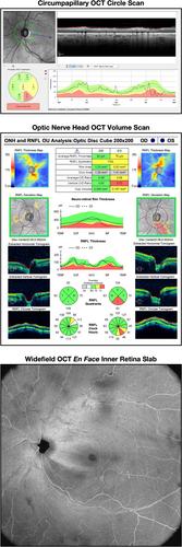
250 eyes from 200 individuals underwent high-resolution microperimetry testing of a hemifield using a stimulus pattern optimised to sample typical arcuate patterns of visual field loss in glaucoma. SAP was performed using a 24–2 stimulus pattern. The presence of glaucomatous damage in each hemifield was subsequently independently assessed by two graders based on a circumpapillary OCT circle scan, 6×6 mm optic disc-centred OCT volume scan, and 15×15 mm widefield OCT volume scan.
Results
The hemifield-based mean total deviation (MTD) and pointwise sensitivity standard deviation (PSD) for high-resolution microperimetry and SAP were both independently associated with the presence of glaucomatous damage in multivariable logistic regression analyses (p ≤ 0.046). Prediction models developed using these two parameters showed significantly higher performance with high-resolution microperimetry compared to SAP (partial area under the receiver operating characteristic curve [pAUC] = 0.86 and 0.75 respectively; p = 0.007). The performance of microperimetry decreased with decreasing resolution when the MTD and PSD values were derived from every second, third, and sixth test locations (pAUC = 0.85 [p = 0.16], 0.85 [p = 0.044] and 0.84 [p < 0.001] respectively), but they were all significantly better than SAP (p ≤ 0.028 for all).
Conclusions
High-resolution microperimetry enabled improved performance for detecting glaucomatous damage compared to SAP, highlighting the potential value of fundus-tracking and higher-resolution sampling for the detection of glaucomatous visual field loss.
期刊介绍:
Clinical & Experimental Ophthalmology is the official journal of The Royal Australian and New Zealand College of Ophthalmologists. The journal publishes peer-reviewed original research and reviews dealing with all aspects of clinical practice and research which are international in scope and application. CEO recognises the importance of collaborative research and welcomes papers that have a direct influence on ophthalmic practice but are not unique to ophthalmology.
 求助内容:
求助内容: 应助结果提醒方式:


