Karim Taha, Youri Bekhuis, Ruben de Bosscher, Christophe Dausin, Marta Orlowska, Ahmed S Youssef, Stéphanie Bézy, Véronique Cornelissen, Lieven Herbots, Rik Willems, Jens-Uwe Voigt, Jan D'hooge, Guido Claessen
{"title":"横波弹性成像揭示运动员和久坐不动的非运动员之间心肌硬度的差异。","authors":"Karim Taha, Youri Bekhuis, Ruben de Bosscher, Christophe Dausin, Marta Orlowska, Ahmed S Youssef, Stéphanie Bézy, Véronique Cornelissen, Lieven Herbots, Rik Willems, Jens-Uwe Voigt, Jan D'hooge, Guido Claessen","doi":"10.1093/ehjimp/qyaf023","DOIUrl":null,"url":null,"abstract":"<p><strong>Aims: </strong>Myocardial stiffening naturally occurs with aging and contributes to diastolic dysfunction. Assessing myocardial stiffness non-invasively could improve the sensitivity of diastolic function evaluation in clinical practice. Shear wave (SW) elastography is a non-invasive tool for quantifying myocardial stiffness, where higher SW velocities indicate increased stiffness. We investigated whether SW elastography could detect differences in myocardial stiffness between athletes and sedentary non-athletes and, during exercise, reveal differences in operational stiffness that may indicate diastolic dysfunction.</p><p><strong>Methods and results: </strong>We enrolled 20 master athletes (median age 60 [IQR 59-66] years) and 17 sedentary non-athletes (median age 58 [IQR 52-71] years). Standard exercise echocardiography revealed no significant differences in diastolic function between the groups. Additionally, ultra-high frame rate imaging was used to measure SW velocities after mitral valve closure (MVC) and aortic valve closure (AVC) at rest and during exercise. At rest, athletes had lower SW velocities after MVC compared to sedentary non-athletes (3.2 ± 0.4 m/s vs. 3.9 ± 0.7 m/s, respectively, <i>P</i> = 0.003). During exercise, SW velocities after AVC significantly increased in sedentary non-athletes but not in athletes (+1.6 ± 1.6 cm/s increase per 1% power output increase vs. 0.0 ± 0.8 cm/s, respectively, <i>P</i> = 0.006). An inverse correlation was found between the increase of SW velocity after AVC during exercise and VO<sub>2</sub>max (<i>r</i> = -0.51, <i>P</i> = 0.003).</p><p><strong>Conclusion: </strong>SW elastography reveals reduced myocardial stiffness in athletes compared to sedentary non-athletes at rest and during exercise, which is not detected by conventional echocardiographic measurements. Exercise-induced changes in SW velocities after AVC may potentially serve as an early marker for detecting diastolic dysfunction.</p>","PeriodicalId":94317,"journal":{"name":"European heart journal. Imaging methods and practice","volume":"2 4","pages":"qyaf023"},"PeriodicalIF":0.0000,"publicationDate":"2025-03-21","publicationTypes":"Journal Article","fieldsOfStudy":null,"isOpenAccess":false,"openAccessPdf":"https://www.ncbi.nlm.nih.gov/pmc/articles/PMC11925635/pdf/","citationCount":"0","resultStr":"{\"title\":\"Shear wave elastography to unmask differences in myocardial stiffness between athletes and sedentary non-athletes.\",\"authors\":\"Karim Taha, Youri Bekhuis, Ruben de Bosscher, Christophe Dausin, Marta Orlowska, Ahmed S Youssef, Stéphanie Bézy, Véronique Cornelissen, Lieven Herbots, Rik Willems, Jens-Uwe Voigt, Jan D'hooge, Guido Claessen\",\"doi\":\"10.1093/ehjimp/qyaf023\",\"DOIUrl\":null,\"url\":null,\"abstract\":\"<p><strong>Aims: </strong>Myocardial stiffening naturally occurs with aging and contributes to diastolic dysfunction. Assessing myocardial stiffness non-invasively could improve the sensitivity of diastolic function evaluation in clinical practice. Shear wave (SW) elastography is a non-invasive tool for quantifying myocardial stiffness, where higher SW velocities indicate increased stiffness. We investigated whether SW elastography could detect differences in myocardial stiffness between athletes and sedentary non-athletes and, during exercise, reveal differences in operational stiffness that may indicate diastolic dysfunction.</p><p><strong>Methods and results: </strong>We enrolled 20 master athletes (median age 60 [IQR 59-66] years) and 17 sedentary non-athletes (median age 58 [IQR 52-71] years). Standard exercise echocardiography revealed no significant differences in diastolic function between the groups. Additionally, ultra-high frame rate imaging was used to measure SW velocities after mitral valve closure (MVC) and aortic valve closure (AVC) at rest and during exercise. At rest, athletes had lower SW velocities after MVC compared to sedentary non-athletes (3.2 ± 0.4 m/s vs. 3.9 ± 0.7 m/s, respectively, <i>P</i> = 0.003). During exercise, SW velocities after AVC significantly increased in sedentary non-athletes but not in athletes (+1.6 ± 1.6 cm/s increase per 1% power output increase vs. 0.0 ± 0.8 cm/s, respectively, <i>P</i> = 0.006). An inverse correlation was found between the increase of SW velocity after AVC during exercise and VO<sub>2</sub>max (<i>r</i> = -0.51, <i>P</i> = 0.003).</p><p><strong>Conclusion: </strong>SW elastography reveals reduced myocardial stiffness in athletes compared to sedentary non-athletes at rest and during exercise, which is not detected by conventional echocardiographic measurements. Exercise-induced changes in SW velocities after AVC may potentially serve as an early marker for detecting diastolic dysfunction.</p>\",\"PeriodicalId\":94317,\"journal\":{\"name\":\"European heart journal. Imaging methods and practice\",\"volume\":\"2 4\",\"pages\":\"qyaf023\"},\"PeriodicalIF\":0.0000,\"publicationDate\":\"2025-03-21\",\"publicationTypes\":\"Journal Article\",\"fieldsOfStudy\":null,\"isOpenAccess\":false,\"openAccessPdf\":\"https://www.ncbi.nlm.nih.gov/pmc/articles/PMC11925635/pdf/\",\"citationCount\":\"0\",\"resultStr\":null,\"platform\":\"Semanticscholar\",\"paperid\":null,\"PeriodicalName\":\"European heart journal. Imaging methods and practice\",\"FirstCategoryId\":\"1085\",\"ListUrlMain\":\"https://doi.org/10.1093/ehjimp/qyaf023\",\"RegionNum\":0,\"RegionCategory\":null,\"ArticlePicture\":[],\"TitleCN\":null,\"AbstractTextCN\":null,\"PMCID\":null,\"EPubDate\":\"2024/10/1 0:00:00\",\"PubModel\":\"eCollection\",\"JCR\":\"\",\"JCRName\":\"\",\"Score\":null,\"Total\":0}","platform":"Semanticscholar","paperid":null,"PeriodicalName":"European heart journal. Imaging methods and practice","FirstCategoryId":"1085","ListUrlMain":"https://doi.org/10.1093/ehjimp/qyaf023","RegionNum":0,"RegionCategory":null,"ArticlePicture":[],"TitleCN":null,"AbstractTextCN":null,"PMCID":null,"EPubDate":"2024/10/1 0:00:00","PubModel":"eCollection","JCR":"","JCRName":"","Score":null,"Total":0}
引用次数: 0
摘要
目的:心肌硬化随着年龄的增长而自然发生,并有助于舒张功能障碍。无创评估心肌僵硬度可提高临床舒张功能评估的敏感性。横波(SW)弹性成像是一种量化心肌刚度的非侵入性工具,较高的横波速度表明心肌刚度增加。我们研究了SW弹性成像是否可以检测运动员和久坐不动的非运动员之间心肌僵硬度的差异,并在运动过程中揭示可能指示舒张功能障碍的操作僵硬度的差异。方法与结果:我们招募了20名运动健将(中位年龄60 [IQR 59-66]岁)和17名久坐不动的非运动员(中位年龄58 [IQR 52-71]岁)。标准运动超声心动图显示各组间舒张功能无显著差异。此外,超高帧率成像用于测量休息和运动时二尖瓣关闭(MVC)和主动脉瓣关闭(AVC)后的SW速度。在休息时,运动员在MVC后的SW速度比久坐的非运动员低(分别为3.2±0.4 m/s和3.9±0.7 m/s, P = 0.003)。运动期间,久坐不动的非运动员在AVC后的SW速度显著增加,而运动员则没有(每1%功率输出增加+1.6±1.6 cm/s vs. 0.0±0.8 cm/s, P = 0.006)。运动期间AVC后SW速度的增加与VO2max呈负相关(r = -0.51, P = 0.003)。结论:SW弹性图显示,与静止不动的非运动员相比,运动员在休息和运动时心肌僵硬度降低,这是传统超声心动图测量无法检测到的。AVC后运动引起的SW速度变化可能作为检测舒张功能障碍的早期标志。
Shear wave elastography to unmask differences in myocardial stiffness between athletes and sedentary non-athletes.
Aims: Myocardial stiffening naturally occurs with aging and contributes to diastolic dysfunction. Assessing myocardial stiffness non-invasively could improve the sensitivity of diastolic function evaluation in clinical practice. Shear wave (SW) elastography is a non-invasive tool for quantifying myocardial stiffness, where higher SW velocities indicate increased stiffness. We investigated whether SW elastography could detect differences in myocardial stiffness between athletes and sedentary non-athletes and, during exercise, reveal differences in operational stiffness that may indicate diastolic dysfunction.
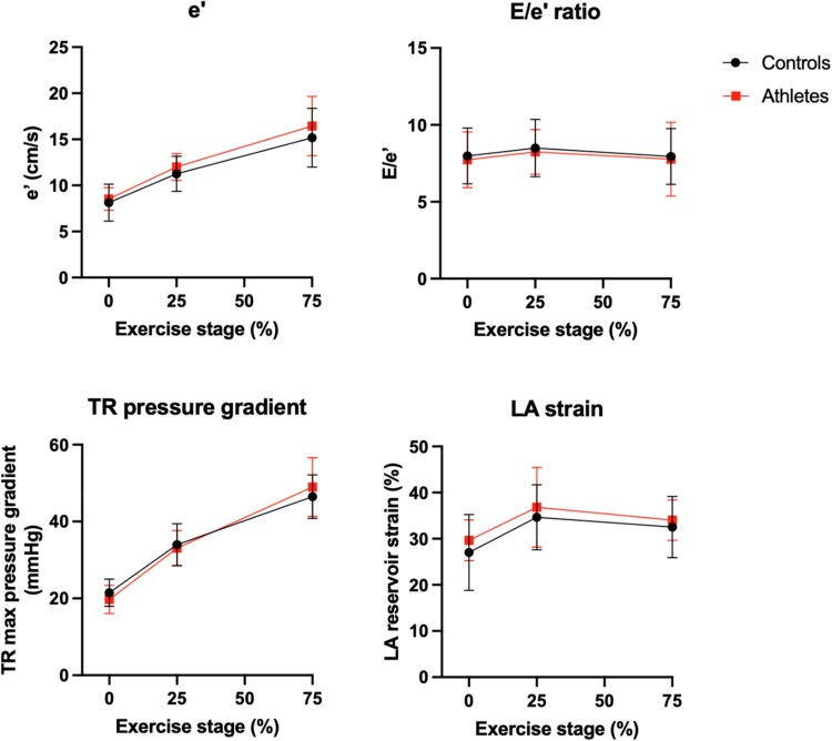
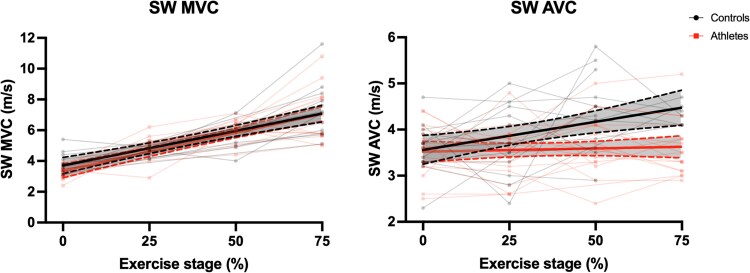
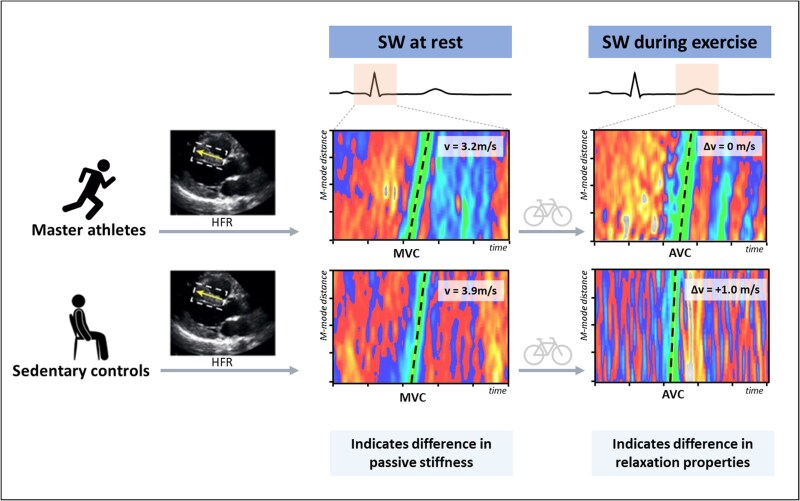
Methods and results: We enrolled 20 master athletes (median age 60 [IQR 59-66] years) and 17 sedentary non-athletes (median age 58 [IQR 52-71] years). Standard exercise echocardiography revealed no significant differences in diastolic function between the groups. Additionally, ultra-high frame rate imaging was used to measure SW velocities after mitral valve closure (MVC) and aortic valve closure (AVC) at rest and during exercise. At rest, athletes had lower SW velocities after MVC compared to sedentary non-athletes (3.2 ± 0.4 m/s vs. 3.9 ± 0.7 m/s, respectively, P = 0.003). During exercise, SW velocities after AVC significantly increased in sedentary non-athletes but not in athletes (+1.6 ± 1.6 cm/s increase per 1% power output increase vs. 0.0 ± 0.8 cm/s, respectively, P = 0.006). An inverse correlation was found between the increase of SW velocity after AVC during exercise and VO2max (r = -0.51, P = 0.003).
Conclusion: SW elastography reveals reduced myocardial stiffness in athletes compared to sedentary non-athletes at rest and during exercise, which is not detected by conventional echocardiographic measurements. Exercise-induced changes in SW velocities after AVC may potentially serve as an early marker for detecting diastolic dysfunction.

 求助内容:
求助内容: 应助结果提醒方式:


