N-G Tzortzakis, S Damaskos, K Dimakopoulou, E Chatzipetros, C Angelopoulos
{"title":"评估种植体周围骨缺损的根尖周射线照相与锥形束 CT 成像:一项体外研究。","authors":"N-G Tzortzakis, S Damaskos, K Dimakopoulou, E Chatzipetros, C Angelopoulos","doi":"10.4317/medoral.26777","DOIUrl":null,"url":null,"abstract":"<p><strong>Background: </strong>Data on the radiographic interpretation of peri-implantitis is still controversial. Thus, our study aimed to: a) investigate the detectability rate of ex-vivo induced peri-implant bone defects (PBDs) between observers using two different imaging methods; Cone Beam Computed Tomography (CBCT) and Periapical Radiographs (PAs), b) investigate the observers' agreement on their ability to detect PBDs according to their level of expertise and, c) determine the sensitivity and specificity of the imaging methods used to detect induced PBDs.</p><p><strong>Material and methods: </strong>Two dried human mandibles were used in which ten dental implants were placed and eight PBDs were created simulating clinical conditions. Radiographic examination using PAs and two CBCT modes [CBCT/N (normal/0.3mm3), and CBCT/HR (HiRes/0.15mm3)] was performed at all experimental stages. All PBDs were recorded for their dimensions using a dental periodontal probe as they were used as a gold standard (GS). Finally, 145 images (49 PAs, 48 CBCT/N, and 48 CBCT/HR) were created and evaluated by nine independent observers. Three oral radiologists (OR), three implantologists (IS), and three general practitioners (GP).</p><p><strong>Results: </strong>PBDs were detected at a higher rate by ORs compared to ISs, and GPs. However, the rate of their agreement, did not reach the nominal level of significance (z-test p-value> 0.05), and also between observers of the same expertise, and between the different imaging methods used: CBCT and PAs (z-test p-value> 0.05). In total, the sensitivity of the CBCTs and PAs method was 95% and 80.5%, respectively. While the specificity for all methods was lower, 57%, 62.2% and 50.4% for CBCT/N, CBCT/H and PAs methods, respectively.</p><p><strong>Conclusions: </strong>Although CBCT performs better than PAs in ex-vivo induced PBDs, further research is needed to evaluate if the present results can be extrapolated to other clinical scenarios and defect configurations.</p>","PeriodicalId":49016,"journal":{"name":"Medicina Oral Patologia Oral Y Cirugia Bucal","volume":" ","pages":"e322-e332"},"PeriodicalIF":2.1000,"publicationDate":"2025-05-01","publicationTypes":"Journal Article","fieldsOfStudy":null,"isOpenAccess":false,"openAccessPdf":"https://www.ncbi.nlm.nih.gov/pmc/articles/PMC12019657/pdf/","citationCount":"0","resultStr":"{\"title\":\"Periapical radiographs vs cone beam CT imaging for the evaluation of peri-implant bone defects: an ex vivo study.\",\"authors\":\"N-G Tzortzakis, S Damaskos, K Dimakopoulou, E Chatzipetros, C Angelopoulos\",\"doi\":\"10.4317/medoral.26777\",\"DOIUrl\":null,\"url\":null,\"abstract\":\"<p><strong>Background: </strong>Data on the radiographic interpretation of peri-implantitis is still controversial. Thus, our study aimed to: a) investigate the detectability rate of ex-vivo induced peri-implant bone defects (PBDs) between observers using two different imaging methods; Cone Beam Computed Tomography (CBCT) and Periapical Radiographs (PAs), b) investigate the observers' agreement on their ability to detect PBDs according to their level of expertise and, c) determine the sensitivity and specificity of the imaging methods used to detect induced PBDs.</p><p><strong>Material and methods: </strong>Two dried human mandibles were used in which ten dental implants were placed and eight PBDs were created simulating clinical conditions. Radiographic examination using PAs and two CBCT modes [CBCT/N (normal/0.3mm3), and CBCT/HR (HiRes/0.15mm3)] was performed at all experimental stages. All PBDs were recorded for their dimensions using a dental periodontal probe as they were used as a gold standard (GS). Finally, 145 images (49 PAs, 48 CBCT/N, and 48 CBCT/HR) were created and evaluated by nine independent observers. Three oral radiologists (OR), three implantologists (IS), and three general practitioners (GP).</p><p><strong>Results: </strong>PBDs were detected at a higher rate by ORs compared to ISs, and GPs. However, the rate of their agreement, did not reach the nominal level of significance (z-test p-value> 0.05), and also between observers of the same expertise, and between the different imaging methods used: CBCT and PAs (z-test p-value> 0.05). In total, the sensitivity of the CBCTs and PAs method was 95% and 80.5%, respectively. While the specificity for all methods was lower, 57%, 62.2% and 50.4% for CBCT/N, CBCT/H and PAs methods, respectively.</p><p><strong>Conclusions: </strong>Although CBCT performs better than PAs in ex-vivo induced PBDs, further research is needed to evaluate if the present results can be extrapolated to other clinical scenarios and defect configurations.</p>\",\"PeriodicalId\":49016,\"journal\":{\"name\":\"Medicina Oral Patologia Oral Y Cirugia Bucal\",\"volume\":\" \",\"pages\":\"e322-e332\"},\"PeriodicalIF\":2.1000,\"publicationDate\":\"2025-05-01\",\"publicationTypes\":\"Journal Article\",\"fieldsOfStudy\":null,\"isOpenAccess\":false,\"openAccessPdf\":\"https://www.ncbi.nlm.nih.gov/pmc/articles/PMC12019657/pdf/\",\"citationCount\":\"0\",\"resultStr\":null,\"platform\":\"Semanticscholar\",\"paperid\":null,\"PeriodicalName\":\"Medicina Oral Patologia Oral Y Cirugia Bucal\",\"FirstCategoryId\":\"3\",\"ListUrlMain\":\"https://doi.org/10.4317/medoral.26777\",\"RegionNum\":3,\"RegionCategory\":\"医学\",\"ArticlePicture\":[],\"TitleCN\":null,\"AbstractTextCN\":null,\"PMCID\":null,\"EPubDate\":\"\",\"PubModel\":\"\",\"JCR\":\"Q2\",\"JCRName\":\"DENTISTRY, ORAL SURGERY & MEDICINE\",\"Score\":null,\"Total\":0}","platform":"Semanticscholar","paperid":null,"PeriodicalName":"Medicina Oral Patologia Oral Y Cirugia Bucal","FirstCategoryId":"3","ListUrlMain":"https://doi.org/10.4317/medoral.26777","RegionNum":3,"RegionCategory":"医学","ArticlePicture":[],"TitleCN":null,"AbstractTextCN":null,"PMCID":null,"EPubDate":"","PubModel":"","JCR":"Q2","JCRName":"DENTISTRY, ORAL SURGERY & MEDICINE","Score":null,"Total":0}
Periapical radiographs vs cone beam CT imaging for the evaluation of peri-implant bone defects: an ex vivo study.
Background: Data on the radiographic interpretation of peri-implantitis is still controversial. Thus, our study aimed to: a) investigate the detectability rate of ex-vivo induced peri-implant bone defects (PBDs) between observers using two different imaging methods; Cone Beam Computed Tomography (CBCT) and Periapical Radiographs (PAs), b) investigate the observers' agreement on their ability to detect PBDs according to their level of expertise and, c) determine the sensitivity and specificity of the imaging methods used to detect induced PBDs.
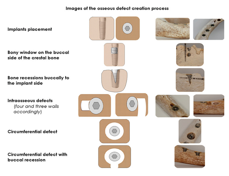
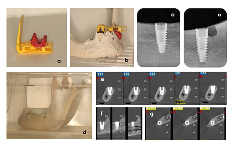
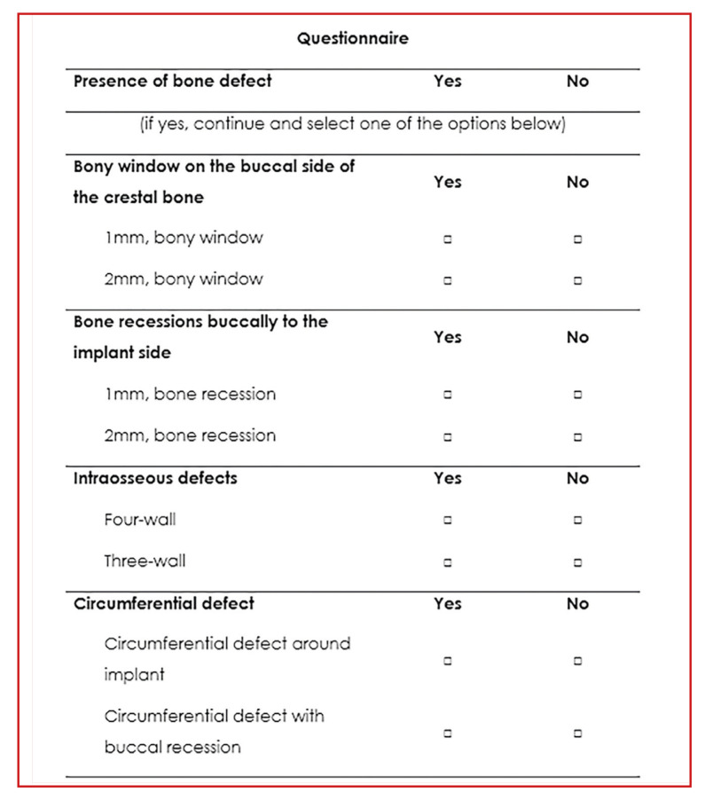
Material and methods: Two dried human mandibles were used in which ten dental implants were placed and eight PBDs were created simulating clinical conditions. Radiographic examination using PAs and two CBCT modes [CBCT/N (normal/0.3mm3), and CBCT/HR (HiRes/0.15mm3)] was performed at all experimental stages. All PBDs were recorded for their dimensions using a dental periodontal probe as they were used as a gold standard (GS). Finally, 145 images (49 PAs, 48 CBCT/N, and 48 CBCT/HR) were created and evaluated by nine independent observers. Three oral radiologists (OR), three implantologists (IS), and three general practitioners (GP).
Results: PBDs were detected at a higher rate by ORs compared to ISs, and GPs. However, the rate of their agreement, did not reach the nominal level of significance (z-test p-value> 0.05), and also between observers of the same expertise, and between the different imaging methods used: CBCT and PAs (z-test p-value> 0.05). In total, the sensitivity of the CBCTs and PAs method was 95% and 80.5%, respectively. While the specificity for all methods was lower, 57%, 62.2% and 50.4% for CBCT/N, CBCT/H and PAs methods, respectively.
Conclusions: Although CBCT performs better than PAs in ex-vivo induced PBDs, further research is needed to evaluate if the present results can be extrapolated to other clinical scenarios and defect configurations.
期刊介绍:
1. Oral Medicine and Pathology:
Clinicopathological as well as medical or surgical management aspects of
diseases affecting oral mucosa, salivary glands, maxillary bones, as well as
orofacial neurological disorders, and systemic conditions with an impact on
the oral cavity.
2. Oral Surgery:
Surgical management aspects of diseases affecting oral mucosa, salivary glands,
maxillary bones, teeth, implants, oral surgical procedures. Surgical management
of diseases affecting head and neck areas.
3. Medically compromised patients in Dentistry:
Articles discussing medical problems in Odontology will also be included, with
a special focus on the clinico-odontological management of medically compromised patients, and considerations regarding high-risk or disabled patients.
4. Implantology
5. Periodontology

 求助内容:
求助内容: 应助结果提醒方式:


