{"title":"额骨毛霉菌病继发角膜穿孔1例。","authors":"Venugopal Anitha, Maneksha Velu, Senthil Babu, Arasi Rajesh, Meenakshi Ravindran","doi":"10.4103/ojo.ojo_221_23","DOIUrl":null,"url":null,"abstract":"<p><p>We present the case of a 52-year-old male with a complex clinical presentation involving frontal sinus osteomyelitis (OM), mechanical ptosis, and extrusion of frontal bone sequestrum under the upper lid skin. He gave a history of chronic sinusitis, an uncontrolled diabetic for the past 2 years. The patient had a history of chronic sinusitis and poorly controlled diabetes for the past 2 years. Histopathological examination of excised sequestrum, stained with hematoxylin and eosin, confirmed a diagnosis of mucormycosis. Anterior segment examination revealed a corneal melt with a shallow anterior chamber. Initial management included the application of an amniotic membrane to promote epithelization. This is the first reported case of frontal bone OM leading to corneal melt, attributed to the mechanical erosion caused by the sequestered bone impinging on the corneal surface.</p>","PeriodicalId":19461,"journal":{"name":"Oman Journal of Ophthalmology","volume":"18 1","pages":"73-76"},"PeriodicalIF":0.0000,"publicationDate":"2025-02-25","publicationTypes":"Journal Article","fieldsOfStudy":null,"isOpenAccess":false,"openAccessPdf":"https://www.ncbi.nlm.nih.gov/pmc/articles/PMC11925373/pdf/","citationCount":"0","resultStr":"{\"title\":\"An unusual case of corneal perforation secondary to Frontal Bone Mucormycosis.\",\"authors\":\"Venugopal Anitha, Maneksha Velu, Senthil Babu, Arasi Rajesh, Meenakshi Ravindran\",\"doi\":\"10.4103/ojo.ojo_221_23\",\"DOIUrl\":null,\"url\":null,\"abstract\":\"<p><p>We present the case of a 52-year-old male with a complex clinical presentation involving frontal sinus osteomyelitis (OM), mechanical ptosis, and extrusion of frontal bone sequestrum under the upper lid skin. He gave a history of chronic sinusitis, an uncontrolled diabetic for the past 2 years. The patient had a history of chronic sinusitis and poorly controlled diabetes for the past 2 years. Histopathological examination of excised sequestrum, stained with hematoxylin and eosin, confirmed a diagnosis of mucormycosis. Anterior segment examination revealed a corneal melt with a shallow anterior chamber. Initial management included the application of an amniotic membrane to promote epithelization. This is the first reported case of frontal bone OM leading to corneal melt, attributed to the mechanical erosion caused by the sequestered bone impinging on the corneal surface.</p>\",\"PeriodicalId\":19461,\"journal\":{\"name\":\"Oman Journal of Ophthalmology\",\"volume\":\"18 1\",\"pages\":\"73-76\"},\"PeriodicalIF\":0.0000,\"publicationDate\":\"2025-02-25\",\"publicationTypes\":\"Journal Article\",\"fieldsOfStudy\":null,\"isOpenAccess\":false,\"openAccessPdf\":\"https://www.ncbi.nlm.nih.gov/pmc/articles/PMC11925373/pdf/\",\"citationCount\":\"0\",\"resultStr\":null,\"platform\":\"Semanticscholar\",\"paperid\":null,\"PeriodicalName\":\"Oman Journal of Ophthalmology\",\"FirstCategoryId\":\"1085\",\"ListUrlMain\":\"https://doi.org/10.4103/ojo.ojo_221_23\",\"RegionNum\":0,\"RegionCategory\":null,\"ArticlePicture\":[],\"TitleCN\":null,\"AbstractTextCN\":null,\"PMCID\":null,\"EPubDate\":\"2025/1/1 0:00:00\",\"PubModel\":\"eCollection\",\"JCR\":\"Q3\",\"JCRName\":\"Medicine\",\"Score\":null,\"Total\":0}","platform":"Semanticscholar","paperid":null,"PeriodicalName":"Oman Journal of Ophthalmology","FirstCategoryId":"1085","ListUrlMain":"https://doi.org/10.4103/ojo.ojo_221_23","RegionNum":0,"RegionCategory":null,"ArticlePicture":[],"TitleCN":null,"AbstractTextCN":null,"PMCID":null,"EPubDate":"2025/1/1 0:00:00","PubModel":"eCollection","JCR":"Q3","JCRName":"Medicine","Score":null,"Total":0}
An unusual case of corneal perforation secondary to Frontal Bone Mucormycosis.
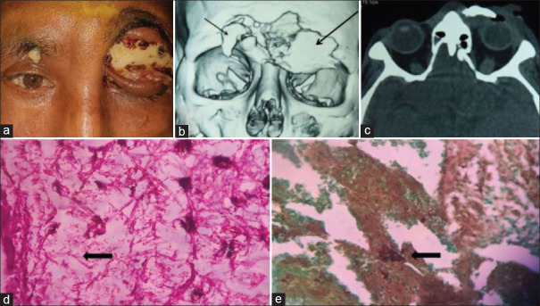
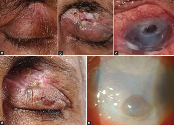
We present the case of a 52-year-old male with a complex clinical presentation involving frontal sinus osteomyelitis (OM), mechanical ptosis, and extrusion of frontal bone sequestrum under the upper lid skin. He gave a history of chronic sinusitis, an uncontrolled diabetic for the past 2 years. The patient had a history of chronic sinusitis and poorly controlled diabetes for the past 2 years. Histopathological examination of excised sequestrum, stained with hematoxylin and eosin, confirmed a diagnosis of mucormycosis. Anterior segment examination revealed a corneal melt with a shallow anterior chamber. Initial management included the application of an amniotic membrane to promote epithelization. This is the first reported case of frontal bone OM leading to corneal melt, attributed to the mechanical erosion caused by the sequestered bone impinging on the corneal surface.
期刊介绍:
To provide a platform for scientific expression of the Oman Ophthalmic Society and the international Ophthalmic community and to provide opportunities for free exchange of ideas and information. To serve as a valuable resource for ophthalmologists, eye-care providers including optometrists, orthoptists, other health care professionals and research workers in all aspects of the field of visual science.

 求助内容:
求助内容: 应助结果提醒方式:


