Selim Doganay, Mehmet Omer Kiristioglu, Gamze Ucan Gunduz
{"title":"一种新颖的手术技术:自体板层巩膜贴片治疗后极创伤性巩膜缺损。","authors":"Selim Doganay, Mehmet Omer Kiristioglu, Gamze Ucan Gunduz","doi":"10.4103/ojo.ojo_104_24","DOIUrl":null,"url":null,"abstract":"<p><p>This case report presents the case of a 60-year-old man who underwent autologous lamellar scleral patch graft (SPG) repair for a large posterior pole scleral defect during pars plana vitrectomy (PPV). Initially, the patient experienced a perforating air rifle pellet injury, necessitating suturing of the entry wound. Six days later, lensectomy and PPV were performed for traumatic cataract, removal of an intraocular foreign body, dense vitreous hemorrhage, and retinal detachment. During PPV, a significant full-thickness scleral defect was found at the posterior pole and repaired using a 3.5 mm × 4.5 mm autologous lamellar SPG sourced from the superotemporal superficial sclera. Postoperatively, the graft fully adhered to the adjacent sclera without complications such as silicone oil leakage or inflammation. This case marks the first instance of utilizing an autologous lamellar SPG for such a large posterior pole defect during PPV, showcasing its effectiveness and safety for such challenging conditions.</p>","PeriodicalId":19461,"journal":{"name":"Oman Journal of Ophthalmology","volume":"18 1","pages":"77-80"},"PeriodicalIF":0.0000,"publicationDate":"2025-02-25","publicationTypes":"Journal Article","fieldsOfStudy":null,"isOpenAccess":false,"openAccessPdf":"https://www.ncbi.nlm.nih.gov/pmc/articles/PMC11925371/pdf/","citationCount":"0","resultStr":"{\"title\":\"A novel surgical technique: \\\"Ab interno\\\" autologous lamellar scleral patch for a large traumatic scleral defect at the posterior pole.\",\"authors\":\"Selim Doganay, Mehmet Omer Kiristioglu, Gamze Ucan Gunduz\",\"doi\":\"10.4103/ojo.ojo_104_24\",\"DOIUrl\":null,\"url\":null,\"abstract\":\"<p><p>This case report presents the case of a 60-year-old man who underwent autologous lamellar scleral patch graft (SPG) repair for a large posterior pole scleral defect during pars plana vitrectomy (PPV). Initially, the patient experienced a perforating air rifle pellet injury, necessitating suturing of the entry wound. Six days later, lensectomy and PPV were performed for traumatic cataract, removal of an intraocular foreign body, dense vitreous hemorrhage, and retinal detachment. During PPV, a significant full-thickness scleral defect was found at the posterior pole and repaired using a 3.5 mm × 4.5 mm autologous lamellar SPG sourced from the superotemporal superficial sclera. Postoperatively, the graft fully adhered to the adjacent sclera without complications such as silicone oil leakage or inflammation. This case marks the first instance of utilizing an autologous lamellar SPG for such a large posterior pole defect during PPV, showcasing its effectiveness and safety for such challenging conditions.</p>\",\"PeriodicalId\":19461,\"journal\":{\"name\":\"Oman Journal of Ophthalmology\",\"volume\":\"18 1\",\"pages\":\"77-80\"},\"PeriodicalIF\":0.0000,\"publicationDate\":\"2025-02-25\",\"publicationTypes\":\"Journal Article\",\"fieldsOfStudy\":null,\"isOpenAccess\":false,\"openAccessPdf\":\"https://www.ncbi.nlm.nih.gov/pmc/articles/PMC11925371/pdf/\",\"citationCount\":\"0\",\"resultStr\":null,\"platform\":\"Semanticscholar\",\"paperid\":null,\"PeriodicalName\":\"Oman Journal of Ophthalmology\",\"FirstCategoryId\":\"1085\",\"ListUrlMain\":\"https://doi.org/10.4103/ojo.ojo_104_24\",\"RegionNum\":0,\"RegionCategory\":null,\"ArticlePicture\":[],\"TitleCN\":null,\"AbstractTextCN\":null,\"PMCID\":null,\"EPubDate\":\"2025/1/1 0:00:00\",\"PubModel\":\"eCollection\",\"JCR\":\"Q3\",\"JCRName\":\"Medicine\",\"Score\":null,\"Total\":0}","platform":"Semanticscholar","paperid":null,"PeriodicalName":"Oman Journal of Ophthalmology","FirstCategoryId":"1085","ListUrlMain":"https://doi.org/10.4103/ojo.ojo_104_24","RegionNum":0,"RegionCategory":null,"ArticlePicture":[],"TitleCN":null,"AbstractTextCN":null,"PMCID":null,"EPubDate":"2025/1/1 0:00:00","PubModel":"eCollection","JCR":"Q3","JCRName":"Medicine","Score":null,"Total":0}
引用次数: 0
摘要
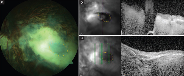
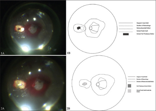
本病例报告介绍了一名 60 岁男性的病例,他在玻璃体旁切除术(PPV)中因后极巩膜大面积缺损而接受了自体片状巩膜补片移植(SPG)修复术。起初,患者的气枪弹丸造成穿孔伤,必须缝合入口伤口。六天后,患者因外伤性白内障、眼内异物取出、玻璃体高密度出血和视网膜脱离而接受了晶状体切除术和 PPV。在 PPV 过程中,发现后极部有明显的全厚巩膜缺损,于是从颞上浅层巩膜取材,用 3.5 mm × 4.5 mm 的自体板层 SPG 进行了修复。术后,移植物与邻近巩膜完全粘连,未出现硅油渗漏或炎症等并发症。该病例标志着在 PPV 手术中首次使用自体板层 SPG 治疗如此大的后极缺损,展示了其在此类挑战性条件下的有效性和安全性。
A novel surgical technique: "Ab interno" autologous lamellar scleral patch for a large traumatic scleral defect at the posterior pole.
This case report presents the case of a 60-year-old man who underwent autologous lamellar scleral patch graft (SPG) repair for a large posterior pole scleral defect during pars plana vitrectomy (PPV). Initially, the patient experienced a perforating air rifle pellet injury, necessitating suturing of the entry wound. Six days later, lensectomy and PPV were performed for traumatic cataract, removal of an intraocular foreign body, dense vitreous hemorrhage, and retinal detachment. During PPV, a significant full-thickness scleral defect was found at the posterior pole and repaired using a 3.5 mm × 4.5 mm autologous lamellar SPG sourced from the superotemporal superficial sclera. Postoperatively, the graft fully adhered to the adjacent sclera without complications such as silicone oil leakage or inflammation. This case marks the first instance of utilizing an autologous lamellar SPG for such a large posterior pole defect during PPV, showcasing its effectiveness and safety for such challenging conditions.
期刊介绍:
To provide a platform for scientific expression of the Oman Ophthalmic Society and the international Ophthalmic community and to provide opportunities for free exchange of ideas and information. To serve as a valuable resource for ophthalmologists, eye-care providers including optometrists, orthoptists, other health care professionals and research workers in all aspects of the field of visual science.

 求助内容:
求助内容: 应助结果提醒方式:


