{"title":"术前计算机断层成像对马颅骨疾病的准确诊断和手术计划:两例先天性畸形报告。","authors":"Takashi Yamaga, Masaaki Tagami, Akiko Takeyama, Fumiki Kato, Tsukasa Suzuki, Masayuki Tagami, Nao Tsuzuki","doi":"10.1294/jes.36.33","DOIUrl":null,"url":null,"abstract":"<p><p>Computed tomography (CT) offers high-resolution, three-dimensional imaging, making it particularly valuable for assessing complex structures, such as the head, especially when conventional radiography and endoscopy are insufficient for a definitive diagnosis. Herein, we present two cases of equine cranial disorders resulting from congenital malformations. In case 1, which had a dentigerous cyst, CT images confirmed the location of the ectopic tooth within the skull and a detailed fistula tube. In case 2, which had temporohyoid osteoarthropathy, CT examination revealed that the stylohyoid bone was malformed, and the inner ear was presumed to be filled with soft tissue with calcification. The information obtained by CT was invaluable, as it allowed for accurate diagnosis and precise surgical planning.</p>","PeriodicalId":35701,"journal":{"name":"Journal of Equine Science","volume":"36 1","pages":"33-37"},"PeriodicalIF":0.0000,"publicationDate":"2025-01-01","publicationTypes":"Journal Article","fieldsOfStudy":null,"isOpenAccess":false,"openAccessPdf":"https://www.ncbi.nlm.nih.gov/pmc/articles/PMC11919546/pdf/","citationCount":"0","resultStr":"{\"title\":\"Preoperative computed tomography imaging for accurate diagnosis and surgical planning in equine cranial disorders: two case reports of congenital malformations.\",\"authors\":\"Takashi Yamaga, Masaaki Tagami, Akiko Takeyama, Fumiki Kato, Tsukasa Suzuki, Masayuki Tagami, Nao Tsuzuki\",\"doi\":\"10.1294/jes.36.33\",\"DOIUrl\":null,\"url\":null,\"abstract\":\"<p><p>Computed tomography (CT) offers high-resolution, three-dimensional imaging, making it particularly valuable for assessing complex structures, such as the head, especially when conventional radiography and endoscopy are insufficient for a definitive diagnosis. Herein, we present two cases of equine cranial disorders resulting from congenital malformations. In case 1, which had a dentigerous cyst, CT images confirmed the location of the ectopic tooth within the skull and a detailed fistula tube. In case 2, which had temporohyoid osteoarthropathy, CT examination revealed that the stylohyoid bone was malformed, and the inner ear was presumed to be filled with soft tissue with calcification. The information obtained by CT was invaluable, as it allowed for accurate diagnosis and precise surgical planning.</p>\",\"PeriodicalId\":35701,\"journal\":{\"name\":\"Journal of Equine Science\",\"volume\":\"36 1\",\"pages\":\"33-37\"},\"PeriodicalIF\":0.0000,\"publicationDate\":\"2025-01-01\",\"publicationTypes\":\"Journal Article\",\"fieldsOfStudy\":null,\"isOpenAccess\":false,\"openAccessPdf\":\"https://www.ncbi.nlm.nih.gov/pmc/articles/PMC11919546/pdf/\",\"citationCount\":\"0\",\"resultStr\":null,\"platform\":\"Semanticscholar\",\"paperid\":null,\"PeriodicalName\":\"Journal of Equine Science\",\"FirstCategoryId\":\"1085\",\"ListUrlMain\":\"https://doi.org/10.1294/jes.36.33\",\"RegionNum\":0,\"RegionCategory\":null,\"ArticlePicture\":[],\"TitleCN\":null,\"AbstractTextCN\":null,\"PMCID\":null,\"EPubDate\":\"2025/3/10 0:00:00\",\"PubModel\":\"Epub\",\"JCR\":\"Q3\",\"JCRName\":\"Veterinary\",\"Score\":null,\"Total\":0}","platform":"Semanticscholar","paperid":null,"PeriodicalName":"Journal of Equine Science","FirstCategoryId":"1085","ListUrlMain":"https://doi.org/10.1294/jes.36.33","RegionNum":0,"RegionCategory":null,"ArticlePicture":[],"TitleCN":null,"AbstractTextCN":null,"PMCID":null,"EPubDate":"2025/3/10 0:00:00","PubModel":"Epub","JCR":"Q3","JCRName":"Veterinary","Score":null,"Total":0}
Preoperative computed tomography imaging for accurate diagnosis and surgical planning in equine cranial disorders: two case reports of congenital malformations.
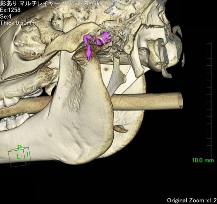
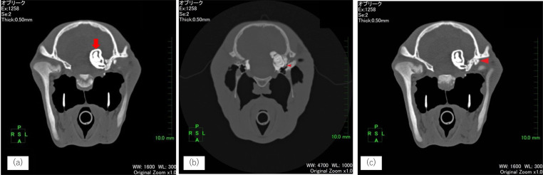
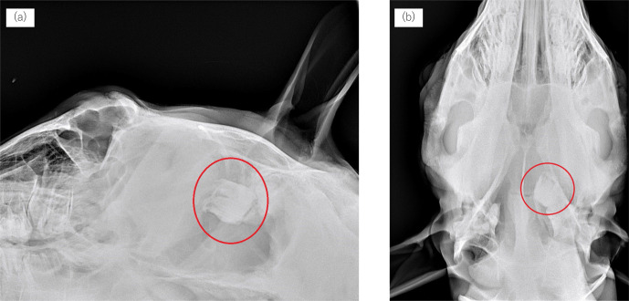
Computed tomography (CT) offers high-resolution, three-dimensional imaging, making it particularly valuable for assessing complex structures, such as the head, especially when conventional radiography and endoscopy are insufficient for a definitive diagnosis. Herein, we present two cases of equine cranial disorders resulting from congenital malformations. In case 1, which had a dentigerous cyst, CT images confirmed the location of the ectopic tooth within the skull and a detailed fistula tube. In case 2, which had temporohyoid osteoarthropathy, CT examination revealed that the stylohyoid bone was malformed, and the inner ear was presumed to be filled with soft tissue with calcification. The information obtained by CT was invaluable, as it allowed for accurate diagnosis and precise surgical planning.

 求助内容:
求助内容: 应助结果提醒方式:


