Chao Lian, Minling He, Chengcheng Zhao, Tianming Wang, Fang Tong, Jianquan Chen, Rong Ju
{"title":"PLIN2:早期非典型子宫内膜增生症的潜在预后标志物。","authors":"Chao Lian, Minling He, Chengcheng Zhao, Tianming Wang, Fang Tong, Jianquan Chen, Rong Ju","doi":"10.3802/jgo.2025.36.e84","DOIUrl":null,"url":null,"abstract":"<p><strong>Objective: </strong>In the background of endometrial hyperplasia triggered by obesity and estrogen, could the perilipin 2 (PLIN2) serve as a possible prognostic marker for early atypical endometrial hyperplasia (AEH)?</p><p><strong>Methods: </strong>A retrospective study examined blood lipid levels in endometrial cancer (EC) or AEH patients. An AEH mice model was established administrating with estradiol and/or high-fat (HF) diet. Hematoxylin and eosin staining were employed to assess pathological changes in the endometrium. Immunohistochemical staining were employed to evaluate the expression of adipose metabolism and endometrial hyperplasia proteins. The Cell Counting Kit-8 assay, cell colony-forming assays, and western blotting were utilized to verify the impact of oleic acid (OA) on HEC-1A cells.</p><p><strong>Results: </strong>The retrospective study revealed elevated blood lipid levels among patients with EC or AEH. Prolonged HF diet stimulated the endometrium to exhibit features of complex atypical hyperplasia. In the early stage, PLIN2 (p=0.006) expression significantly increased with endometrial glandular hyperplasia. Both PLIN2 (p=0.008) and progesterone receptor (PR; p=0.019) exhibited elevated expression concurrent with simple endometrial hyperplasia. When AEH occurred, there were notable rise in the expression of PLIN2 (p<0.001), PR (p=0.044), and estrogen receptor (p=0.045). The atypical hyperplasia glands demonstrated notably elevated PLIN2 expression in comparison to surrounding normal glands in AEH lesions. OA was found to enhance the proliferation and clonal formation of HEC-1A cells. HEC-1A cells induced by OA demonstrated elevated autophagy levels accompanied by enhanced expression of PLIN2.</p><p><strong>Conclusion: </strong>PLIN2 may potentially serve as a biomarker for early development of AEH and EC, facilitating diagnosis and intervention and contributing to the determination of prognosis.</p>","PeriodicalId":15868,"journal":{"name":"Journal of Gynecologic Oncology","volume":" ","pages":"e84"},"PeriodicalIF":3.7000,"publicationDate":"2025-09-01","publicationTypes":"Journal Article","fieldsOfStudy":null,"isOpenAccess":false,"openAccessPdf":"https://www.ncbi.nlm.nih.gov/pmc/articles/PMC12426750/pdf/","citationCount":"0","resultStr":"{\"title\":\"PLIN2: a potential prognostic markers of early-stage atypical endometrial hyperplasia.\",\"authors\":\"Chao Lian, Minling He, Chengcheng Zhao, Tianming Wang, Fang Tong, Jianquan Chen, Rong Ju\",\"doi\":\"10.3802/jgo.2025.36.e84\",\"DOIUrl\":null,\"url\":null,\"abstract\":\"<p><strong>Objective: </strong>In the background of endometrial hyperplasia triggered by obesity and estrogen, could the perilipin 2 (PLIN2) serve as a possible prognostic marker for early atypical endometrial hyperplasia (AEH)?</p><p><strong>Methods: </strong>A retrospective study examined blood lipid levels in endometrial cancer (EC) or AEH patients. An AEH mice model was established administrating with estradiol and/or high-fat (HF) diet. Hematoxylin and eosin staining were employed to assess pathological changes in the endometrium. Immunohistochemical staining were employed to evaluate the expression of adipose metabolism and endometrial hyperplasia proteins. The Cell Counting Kit-8 assay, cell colony-forming assays, and western blotting were utilized to verify the impact of oleic acid (OA) on HEC-1A cells.</p><p><strong>Results: </strong>The retrospective study revealed elevated blood lipid levels among patients with EC or AEH. Prolonged HF diet stimulated the endometrium to exhibit features of complex atypical hyperplasia. In the early stage, PLIN2 (p=0.006) expression significantly increased with endometrial glandular hyperplasia. Both PLIN2 (p=0.008) and progesterone receptor (PR; p=0.019) exhibited elevated expression concurrent with simple endometrial hyperplasia. When AEH occurred, there were notable rise in the expression of PLIN2 (p<0.001), PR (p=0.044), and estrogen receptor (p=0.045). The atypical hyperplasia glands demonstrated notably elevated PLIN2 expression in comparison to surrounding normal glands in AEH lesions. OA was found to enhance the proliferation and clonal formation of HEC-1A cells. HEC-1A cells induced by OA demonstrated elevated autophagy levels accompanied by enhanced expression of PLIN2.</p><p><strong>Conclusion: </strong>PLIN2 may potentially serve as a biomarker for early development of AEH and EC, facilitating diagnosis and intervention and contributing to the determination of prognosis.</p>\",\"PeriodicalId\":15868,\"journal\":{\"name\":\"Journal of Gynecologic Oncology\",\"volume\":\" \",\"pages\":\"e84\"},\"PeriodicalIF\":3.7000,\"publicationDate\":\"2025-09-01\",\"publicationTypes\":\"Journal Article\",\"fieldsOfStudy\":null,\"isOpenAccess\":false,\"openAccessPdf\":\"https://www.ncbi.nlm.nih.gov/pmc/articles/PMC12426750/pdf/\",\"citationCount\":\"0\",\"resultStr\":null,\"platform\":\"Semanticscholar\",\"paperid\":null,\"PeriodicalName\":\"Journal of Gynecologic Oncology\",\"FirstCategoryId\":\"3\",\"ListUrlMain\":\"https://doi.org/10.3802/jgo.2025.36.e84\",\"RegionNum\":2,\"RegionCategory\":\"医学\",\"ArticlePicture\":[],\"TitleCN\":null,\"AbstractTextCN\":null,\"PMCID\":null,\"EPubDate\":\"2025/3/4 0:00:00\",\"PubModel\":\"Epub\",\"JCR\":\"Q1\",\"JCRName\":\"OBSTETRICS & GYNECOLOGY\",\"Score\":null,\"Total\":0}","platform":"Semanticscholar","paperid":null,"PeriodicalName":"Journal of Gynecologic Oncology","FirstCategoryId":"3","ListUrlMain":"https://doi.org/10.3802/jgo.2025.36.e84","RegionNum":2,"RegionCategory":"医学","ArticlePicture":[],"TitleCN":null,"AbstractTextCN":null,"PMCID":null,"EPubDate":"2025/3/4 0:00:00","PubModel":"Epub","JCR":"Q1","JCRName":"OBSTETRICS & GYNECOLOGY","Score":null,"Total":0}
PLIN2: a potential prognostic markers of early-stage atypical endometrial hyperplasia.
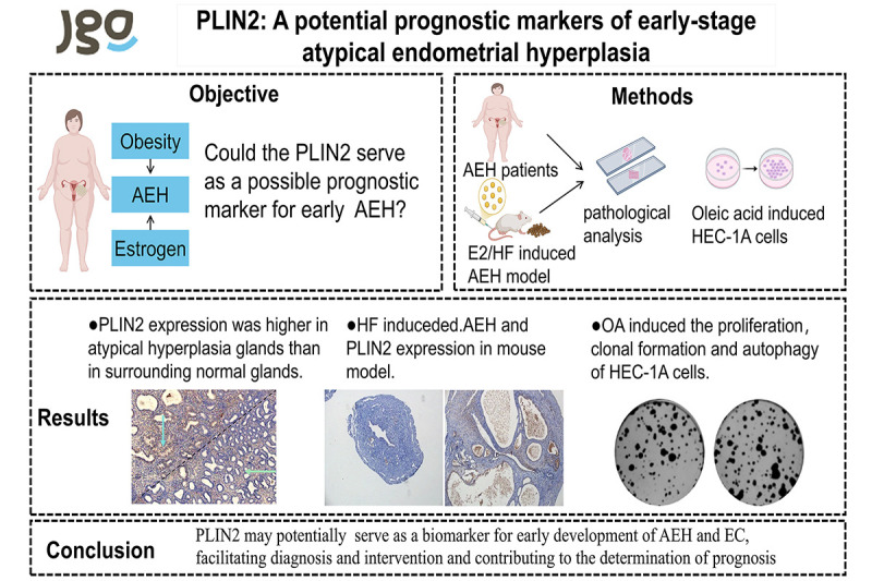
Objective: In the background of endometrial hyperplasia triggered by obesity and estrogen, could the perilipin 2 (PLIN2) serve as a possible prognostic marker for early atypical endometrial hyperplasia (AEH)?
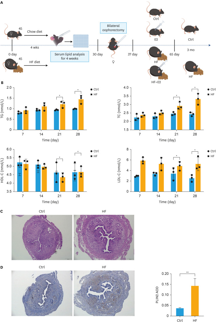
Methods: A retrospective study examined blood lipid levels in endometrial cancer (EC) or AEH patients. An AEH mice model was established administrating with estradiol and/or high-fat (HF) diet. Hematoxylin and eosin staining were employed to assess pathological changes in the endometrium. Immunohistochemical staining were employed to evaluate the expression of adipose metabolism and endometrial hyperplasia proteins. The Cell Counting Kit-8 assay, cell colony-forming assays, and western blotting were utilized to verify the impact of oleic acid (OA) on HEC-1A cells.
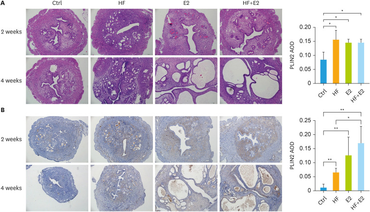
Results: The retrospective study revealed elevated blood lipid levels among patients with EC or AEH. Prolonged HF diet stimulated the endometrium to exhibit features of complex atypical hyperplasia. In the early stage, PLIN2 (p=0.006) expression significantly increased with endometrial glandular hyperplasia. Both PLIN2 (p=0.008) and progesterone receptor (PR; p=0.019) exhibited elevated expression concurrent with simple endometrial hyperplasia. When AEH occurred, there were notable rise in the expression of PLIN2 (p<0.001), PR (p=0.044), and estrogen receptor (p=0.045). The atypical hyperplasia glands demonstrated notably elevated PLIN2 expression in comparison to surrounding normal glands in AEH lesions. OA was found to enhance the proliferation and clonal formation of HEC-1A cells. HEC-1A cells induced by OA demonstrated elevated autophagy levels accompanied by enhanced expression of PLIN2.
Conclusion: PLIN2 may potentially serve as a biomarker for early development of AEH and EC, facilitating diagnosis and intervention and contributing to the determination of prognosis.
期刊介绍:
The Journal of Gynecologic Oncology (JGO) is an official publication of the Asian Society of Gynecologic Oncology. Abbreviated title is ''J Gynecol Oncol''. It was launched in 1990. The JGO''s aim is to publish the highest quality manuscripts dedicated to the advancement of care of the patients with gynecologic cancer. It is an international peer-reviewed periodical journal that is published bimonthly (January, March, May, July, September, and November). Supplement numbers are at times published. The journal publishes editorials, original and review articles, correspondence, book review, etc.
 求助内容:
求助内容: 应助结果提醒方式:


